
Corneal Ulcer: Symptoms, Diagnosis, Treatment
The cornea is the transparent dome at the front of the eye through which light passes. When a deep, inflamed defect forms, it's called a corneal ulcer. This type of damage is a condition where time often counts.
The danger is that a deep defect leaves a scar. This permanently reduces vision. Therefore, a corneal ulcer requires more than just waiting for it to heal. An urgent examination by a specialist is necessary. Restoring the transparency of this membrane is difficult, and sometimes impossible.
Below, we'll discuss the causes and symptoms of corneal ulcers. We'll show you how to treat them, from the initial examination to monitoring their healing. The sooner treatment is started, the higher the chance of preserving clarity and visual acuity.

specialists

equipment

treatment






Causes and risk factors

There are several causes, and treatment directly depends on them. Most often, the defect is triggered by infection, but it is not the only one.
Infections, microtraumas, and inflammation
The most common mechanism is a corneal infection: bacteria, herpes viruses, and, less commonly, fungi or protozoa. Microtraumas, such as scratches from a twig, a grain of sand, or the edge of a lens, open the door for microbes. Bacterial keratitis develops rapidly in these conditions, within days, sometimes even hours.
Contact lenses and poor hygiene
Contact lenses are a separate risk factor, especially if worn overnight, infrequently replaced, or poorly maintained. A warm, moist environment develops under the lens, which is conducive to microbes. Therefore, at the first sign of discomfort, remove it and do not put it back on until an examination.
Dry eye, injuries, concomitant diseases
Dry eye surface, burns, previous herpes, eyelid diseases, and a weak immune system undermine the eye's defenses. Under these conditions, even minor injuries heal more slowly and can more easily develop into ulcers. Some patients experience a combination of these conditions.
How we conduct diagnostics
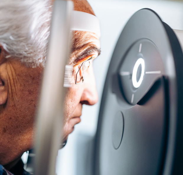
Corneal ulcer diagnosis begins with a detailed examination and takes little time. The goal is to determine the depth of the defect and its cause, as this determines all subsequent treatment.
Slit Lamp Examination
Under an illuminated microscope—a slit lamp—the doctor assesses the size, depth, and edges of the defect, as well as the condition of the anterior chamber. This is the primary method of initial assessment, providing immediate clarity.
Fluorescein Test and Corneal Defect Assessment
A drop of fluorescein dye stains the damaged area, making the defect clearly visible under blue light. The test shows the precise boundaries of the ulcer and allows for monitoring healing from visit to visit.
Additional tests if infection is suspected
If the nature of the inflammation is unclear, a corneal scraping is taken for microbiological analysis. This identifies the pathogen and its sensitivity to medications, which determines the accuracy of treatment, especially in persistent cases.
Symptoms of a corneal ulcer
The symptoms are usually intense. The eye reacts with pain and doesn't let you forget it for a minute.
Pain, redness, and tearing
The main symptoms are sharp pain in the eye, severe redness, and profuse tearing. The discomfort intensifies with each blink, and a reflexive urge to close the eye is felt.
Photophobia and Foreign Body Sensation
Photophobia and a persistent foreign body sensation, as if a speck of dust were lodged under the eyelid, follow. The eye urges to close and shield it from the bright light, and trying to look closely intensifies the stinging.
Corneal Clouding and Decreased Vision
As the defect deepens, corneal clouding appears. Vision deteriorates, ranging from a slight haze to a noticeable decrease in acuity. This symptom indicates involvement of the deep layers of the cornea and requires special vigilance.
Below is a brief list of warning signs. If at least some of these match, an appointment should not be delayed.
- Severe eye pain
- Photophobia
- Corneal Clouding
- Decreased Vision
- Purulent Discharge or Severe Redness
None of these symptoms resolve on their own. Each is a reason for urgent medical attention.
General information
Corneal Ulcer Treatment: How We Help Patients
Corneal ulcer treatment focuses on the cause, not just the symptoms. The treatment plan depends on the depth of the lesion and the type of pathogen.
Treatment Selection Based on the Cause of the Disease
For bacterial causes, intensive antibacterial drops are prescribed, for herpes, antiviral agents, and for fungal infections, antifungal agents. Hormonal medications are not used without medical advice, as they can worsen the defect. All treatment is supervised by a physician, and the frequency of instillations during the acute phase is sometimes increased to several times an hour.
Healing Monitoring and Regimens Adjustment
During the acute phase, the patient is examined frequently. The specialist monitors whether the defect is improving and adjusts the treatment plan if necessary. This monitoring prevents the process from getting out of control and detects complications early.
Monitoring until the corneal surface is restored
Therapy continues until complete epithelialization—the healing of the surface. Even after the pain has subsided, an examination is necessary to assess the strength of the healed area and the transparency of the cornea. Cloudiness may remain at the site of a deep defect, and this is discussed separately with the patient.




Frequently Asked Questions
Can a corneal ulcer be cured without surgery?
How long does it take for the cornea to heal?
When should I seek urgent attention?
Can I wear contact lenses after treatment?
Our doctors





















This award is given to clinics with the highest ratings according to user ratings, a large number of requests from this site, and in the absence of critical violations.

This award is given to clinics with the highest ratings according to user ratings. It means that the place is known, loved, and definitely worth visiting.

The ProDoctors portal collected 500 thousand reviews, compiled a rating of doctors based on them and awarded the best. We are proud that our doctors are among those awarded.
Make an appointment at a convenient time on the nearest date
Price
Other services

Appointment to the doctor
Reviews





















Our clinics


Application “Personal Account K+31”












What is a corneal ulcer and why is it dangerous?
This membrane consists of several layers. The superficial epithelium covers the eye from the outside, and underneath it lies the stroma, which is responsible for strength and transparency. Ulceration destroys these layers, causing inflammation at the site of the defect, which can extend deeper.
What does a corneal lesion look like?
This type of corneal lesion is noticeable as a cloudy spot or depression on the transparent membrane, around which the eye turns red. Sometimes pus accumulates in the anterior chamber, and a white layer is visible at the bottom. The person notices the whitish spot and feels increasing cloudiness.
Why does this condition require urgent examination?
The defect deepens quickly, especially with infection. Delay can lead to scarring, and in severe cases, to perforation of the membrane and penetration of infection into the eye. This is why corneal ulcers are considered an emergency in ophthalmology, along with injuries and burns.