Eyeball Ultrasound (B-scan, eye echography) in Moscow

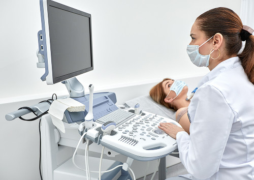
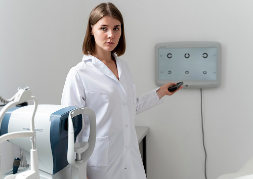
When a doctor needs to assess the internal condition of the visual organs, high-tech ultrasound comes to the rescue. In our medical practice, eyeball ultrasound is recognized as one of the most informative and fastest ways to visualize the internal structures of the eye. This method uses ultrasound waves that reflect off tissues of varying densities, forming a detailed two-dimensional image on the device's screen.

Eyeball ultrasound is available at the K31 Clinic in Moscow.

Best specialists
Best
specialists
Expert equipment
Expert
equipment
Advanced diagnostic treatment
Advanced diagnostic
treatment
Download the application “Personal Account K+31”
Everything about your health in one click!
Subscribe to social networks K+31
To keep up to date with current offers and receive useful advice about your health.

When is an ultrasound of the eyeball prescribed?

There are a number of subjective manifestations for which we recommend that patients immediately undergo an ultrasound examination.

Symptoms that require an eye examination

A doctor may order a B-scan of the eye in the following situations:

  • Sudden decrease in vision or complete loss of vision without apparent external cause
  • Flashes, lightning, and floating spots before the eyes
  • Eye trauma of any severity and location
  • Suspected dangerous condition such as retinal detachment
  • Vitreous hemorrhage that interferes with normal vision
  • Inability to examine the fundus due to clouding of the optical media

When a routine examination is not enough

In ophthalmology, a standard fundus examination is performed using a light beam and special lenses, but this method only works when all internal media of the eye are completely transparent.

A standard diagnosis is impossible if the patient:

  • A mature cataract is developing
  • A significant corneal opacity is observed
  • A massive intraocular hemorrhage has occurred

In this case, the light beam simply cannot penetrate the posterior segment of the eye. Ocular ultrasound becomes an indispensable tool, as ultrasound waves easily penetrate any opacities in the ocular media and allow for a detailed examination of the deep layers.

After injuries, hemorrhages, and surgeries

Ultrasound B-scanning of the eye allows us to quickly assess the extent of damage, check the integrity of the endometrium, and rule out hidden ruptures. This method is also extremely important in the postoperative period for monitoring the position of the artificial lens, assessing the condition of the endometrium after major vitreoretinal procedures, and monitoring the dynamics of blood resorption.

What does a B-scan of the eye show?

Vitreous Body and Transparent Media of the Eye

During this examination, we are able to examine in detail the gel-like substance that fills most of the optic cup. Normally, it should be completely transparent and homogeneous.

Ultrasound imaging allows us to detect changes such as destruction or degenerative opacification of the vitreous body, determine the presence of inflammatory exudates, and identify thin connective tissue strands that can cause dangerous damage to adjacent structures.

Retina, Choroid, and Optic Nerve

Using ultrasound, we assess the topography and thickness of the membranes, as well as the condition of the optic disc and retrobulbar space. The device's screen displays any changes in the shape of the optic cup, signs of tissue edema, congestion in the nerve area, or localized membrane delamination.

Retinal detachment, hemorrhages, foreign bodies

Timely retinal diagnosis is critical when there is a risk of vision loss, and ultrasound is ideal for this task. The image clearly visualizes retinal detachment, allowing us to determine its exact height, extent, and the presence of fixed folds. The examination also accurately detects vitreous hemorrhage, which appears on the monitor as a cluster of multiple mobile opacities of varying density. If a patient has suffered an injury, ultrasound can quickly detect a radiolucent foreign body in the eye, establish its precise coordinates, and determine the extent of damage for planning emergency surgery.

Large lesions and post-traumatic changes

Ultrasound can detect the presence of benign or malignant tumors, measure their precise dimensions in all projections, and assess the growth pattern and extent of invasion into adjacent tissue. When examining the consequences of old injuries, ultrasound can reveal areas of fibrosis, the presence of scars, and signs of wrinkling or subatrophy of the eyeball.

General information

How is an eye ultrasound performed?

Is any preparation necessary?

No, it is not. The only recommendation for women is to completely avoid wearing eye makeup on the day of the examination. If you regularly wear contact lenses, you will need to remove them.

Procedure Steps

The eye ultrasound itself is performed in the comfortable and calm environment of the examination room. The patient sits in a chair or lies down on a table, after which the doctor explains all the stages of the examination in detail. The procedure can be performed in two ways:

  • Through closed eyelids
  • Through the corneal surface after preliminary anesthetic drops have been instilled.

The most common and comfortable method is through a closed eyelid. A small amount of gel is applied to the skin of the eyelid. The doctor places the transducer on the eyelid and moves it in different directions, asking the patient to look up, down, or sideways.

How long does the examination last?

Typically, an examination of one eye takes no more than 5-10 minutes, and a comprehensive ultrasound of the eyeball (B-scan) for both eyes takes about 15 minutes. The doctor requires a few more minutes to analyze the resulting echograms, measure all necessary parameters, and prepare an official medical report.

How safe and comfortable is it?

This is a safe diagnostic method. Ultrasound waves do not damage living tissue, so the examination can be performed an unlimited number of times. The patient experiences no pain or significant discomfort while the transducer is in contact with the skin.

How is an eye ultrasound performed?

B-scan and other diagnostic methods: what's the difference?

How is it different from OCT?

Optical coherence tomography (OCT) uses a light beam, which allows the layers of the retina to be viewed at the cellular level, but light cannot penetrate the cloudy environment of the eye. An ultrasound B-scan uses a sound wave, which has much greater penetrating power and allows for a general anatomical picture to be seen even when the inside of the eye is filled with blood or the lens has completely lost its transparency.

How is it different from a standard ophthalmological examination?

A standard ophthalmological examination using lenses and lamps allows the doctor to evaluate only the visible surface of the retina and the condition of the blood vessels in the fundus, provided they are accessible to the light beam. Ultrasound allows us to assess the condition of the tissues behind the eyeball and measure the precise linear dimensions of internal structures.

For a better understanding of the differences between the main methods, we have compiled a comparative table:

Method What it helps to assess When it is used
Ultrasound of the eyeball (B-scan) Internal structures of the eye, vitreous body, retina If examination is difficult, after injuries, if detachment is suspected
OCT Retinal layers and optic nerve at the micro level For a detailed assessment of the macula and retina in transparent Media
Ophthalmoscopy Visible fundus and large vessels When the ocular media are sufficiently transparent for preventive monitoring

When methods complement each other

We never oppose these diagnostic methods, but rather use them in intelligent synergy. For example, when preparing a patient for cataract surgery, ultrasound is necessary to rule out gross pathology in the posterior pole of the eye, and after successful lens replacement and restoration of medial transparency, we prescribe OCT for a detailed analysis of the central retinal zone.

B-scan and other diagnostic methods: what's the difference?

Why patients choose us

Experienced ophthalmologists

Examinations are performed by highly qualified ophthalmologists who have undergone specialized training in ophthalmological ultrasound diagnostics and have extensive practical experience.

Modern equipment and accurate interpretation

Our diagnostic room is equipped with the latest generation of advanced ultrasound systems. This high-resolution equipment allows us to obtain clear, high-contrast images of the internal structures of the eye with minimal interference.

Clear conclusion and further recommendations

After the scan, the doctor will explain your eye condition in detail, show you key changes on the monitor screen, and explain each line of the report.

Quick appointments and convenient appointment format

We offer quick appointments at convenient times. You can undergo a comprehensive ophthalmological examination and receive results on the same day of your visit, which is especially important if acute symptoms develop.

Why patients choose us

How to schedule an eye ultrasound in Moscow

When is it best not to postpone an examination?

If you experience a sudden decrease in vision, a dark cloud appears in front of your eye, or after a strong blow, you should schedule an eye examination immediately.

What to bring to the appointment?

Please bring your passport to complete your medical record and the results of all previous ophthalmologist examinations or discharge summaries from previous eye surgeries, if available.

What to do after receiving the results?

The report itself is not a final diagnosis. Immediately after the procedure, you will be offered a consultation with an ophthalmologist, who will compare the B-scan data with the results of other tests, establish an accurate diagnosis, and prescribe treatment.

How to schedule an eye ultrasound in Moscow

Frequently Asked Questions

What does an eye ultrasound (B-scan) reveal?

We use this method to visualize changes in the eye and identify hemorrhages, retinal detachments, foreign bodies, and other pathological changes.

Is a B-scan painful?

No, it's a non-invasive and usually comfortable examination. The procedure is quick and doesn't cause significant discomfort.

When should you schedule an urgent eye ultrasound?

This examination is especially important after an eye injury, if there is sudden vision loss, flashes, floaters, or fog, or if there is a suspicion of retinal hemorrhage or detachment.

Sign up for a consultation

Sign up for a consultation

An ultrasound scan is a simple, accessible, and completely painless way to ensure the safety of your eye's internal structures or to begin treating disease early.

"In practice, we most often prescribe a B-scan when a standard examination doesn't provide a complete picture. This helps us quickly assess the condition of the eye's internal structures and choose a course of action without wasting time."

We will conduct an eye examination in a comfortable environment, provide comprehensive answers to all your questions, and offer effective solutions for preserving your vision.

Take a confident step toward maintaining your eye health—schedule an eye ultrasound (B-scan) in Moscow by filling out the form on our website or by contacting us by phone and receive expert diagnostics from leading specialists.

Our doctors

Karapetyan Arevik Samvelovna
Experience 12 years
Make an appointment
Karapetyan
Arevik Samvelovna
Ophthalmologist, Laser Surgeon
Batalina Larisa Vladimirovna
Experience 33 years
Make an appointment
Batalina
Larisa Vladimirovna
Deputy chief physician for ophthalmology, ophthalmic surgeon, leading specialist in laser vision correction
Amiryan Anush Gamletovna
Experience 28 years
Make an appointment
Amiryan
Anush Gamletovna
Ophthalmic oncologist surgeon
Medvedev Yulia Alexandrovna
Experience 11 years
Make an appointment
Medvedev
Yulia Alexandrovna
Ophthalmologist, Laser Surgeon
Rudkovskaya Elena Mikhailovna
Experience 38 years
Make an appointment
Rudkovskaya
Elena Mikhailovna
Ophthalmologist
Tegniryadnova Ekaterina Valerievna
Experience 20 years
Make an appointment
Tegniryadnova
Ekaterina Valerievna
Ophthalmologist, laser surgeon
Fadeeva Victoria Anatolievna
Experience 13 years
Make an appointment
Fadeeva
Victoria Anatolievna
Doctor ophthalmologist, laser surgeon
Shamsetdinova Leila Tagirovna
Experience 17 years
Make an appointment
Shamsetdinova
Leila Tagirovna
Pediatric ophthalmologist
Gavrilina Polina Dmitrievna
Experience 8 years
Make an appointment
Gavrilina
Polina Dmitrievna
Ophthalmologist, laser surgeon
Sugonyaeva Olga Yurievna
Experience 26 years
Make an appointment
Sugonyaeva
Olga Yurievna
Ophthalmologist, laser surgeon
Akhiyarova Azalia Azatovna
Experience 4 years
Make an appointment
Akhiyarova
Azalia Azatovna
Ophthalmologist
Putintseva Polina Andreevna
Experience 6 years
Make an appointment
Putintseva
Polina Andreevna
Ophthalmologist
Agafonova Oksana Sergeevna
Experience 21 year
Make an appointment
Agafonova
Oksana Sergeevna
Ophthalmologist, pediatric
Kazennov Alexey Nikolaevich
Experience 19 years
Make an appointment
Kazennov
Alexey Nikolaevich
Ophthalmologist, ophthalmologist-surgeon
Petrova Maria Grigorievna
Experience 14 years
Make an appointment
Petrova
Maria Grigorievna
Ophthalmologist, ophthalmic surgeon
Mazmanyan Karen Akopovich
Experience 28 years
Make an appointment
Mazmanyan
Karen Akopovich
Ophthalmic surgeon
Suchkova Valeria Alekseevna
Experience 6 years
Make an appointment
Suchkova
Valeria Alekseevna
Ophthalmologist
Khubieva Salima Aslanbievna
Experience 7 years
Make an appointment
Khubieva
Salima Aslanbievna
Ophthalmologist, laser surgeon
Rytik Nina Petrovna
Experience 15 years
Make an appointment
Rytik
Nina Petrovna
Ophthalmologist
All specialists
2GIS Award
2GIS Award

This award is given to clinics with the highest ratings according to user ratings, a large number of requests from this site, and in the absence of critical violations.

«Good place» according to Yandex

This award is given to clinics with the highest ratings according to user ratings. It means that the place is known, loved, and definitely worth visiting.

Our doctors are laureates of the ProDoctors Award

The ProDoctors portal collected 500 thousand reviews, compiled a rating of doctors based on them and awarded the best. We are proud that our doctors are among those awarded.

Make an appointment at a convenient time on the nearest date

Price

Reception
Price
Initial Ophthalmologist Appointment
from 6 450 ₽
Corneal Collagen Crosslinking with Ultraviolet Irradiation
from 52 500 ₽
Femtosecond Laser-Assisted Intrastromal Segment Implantation
from 200 000 ₽

Other services

Laser vision correction Phacoemulsification of cataract with IOL implantation of any complexity Vitreoretinal surgeries of any complexity (vitrectomy) Laser photocoagulation of the retina during tears and degenerative changes in the retina Laser photocoagulation for diabetic retinopathy Intravitreal administration of Anti-VEGF drugs, hormonal implants Antiglaucomatous operations using various drains YAG laser dissection of the posterior capsule for secondary cataract Cataract treatment Treatment of glaucoma Retinal dystrophic changes Vitreous pathology Injury to the eye Post-traumatic and postoperative complications Myopia treatment Farsightedness Astigmatism Retinal disinsertion Non-contact laser procedure SMARTSURFACE Keratoplasty Femto SUPER LASIK Laser coagulation of the retina of the eye treatment on Ellex Integre Pro Scan Treatment of the retina with the Navilas laser system Femtolaser Cataract Removal Selection of children's night lenses Laser vision correction Clear Офтальмолог-онколог в Москве Treatment of keratoconus Laser coagulation of the retina Refractometry Retrobulbar injections Restoration of vision Lacrimal ducts Eye wash Synoptophore Forbis ESOM Pediatric ophthalmologist Ophthalmologist Glasses Biomicroscopy Night lenses Photo of an eye Eyelid suturing Selection of lenses Retinal laser Vision test Vitrectomy Vitrectomy Vitrectomy Eye pressure Ophthalmoscopy Fundus Gonioscopy Discision Orbitotomy Ophthalmodiagnostics OCT of the optic disc Keratoplasty Crystalline lens replacement Lacrimal duct Eye biometrics Angle of strabismus Lacrimal point IOL implantation Heterophoria Refraction Needling pillows Sclerectomy Micropulse laser Eyelid massage Intravitreal injections Iridoplasty Accommodation Bacterial culture Ophthalmology Cyclophotocoagulation Laser trabeculoplasty Laser stimulation Makdel Iridotomy Goniopuncture Corneal crosslinking Perimetry Pachymetry Angio-OCT Tubular test Color vision Convergence test Autofluorescence Eye mobility Corneal diagnostics Corneal transplant Skiascopy Strabismus Injection in the eye Tonometry Schirmer's test Trabeculotomy Diaphanoscopy Foreign body Foreign body Eyelid neoplasms Retinal tear Pterygium Chalazion Pneumatic retinopexy Glaucoma Eye biometrics Improving vision Cataract removal An attack of glaucoma Night lenses Eyelid ptosis Vision correction Angiography of the eye Retinoblastoma Laser coagulation PRK Orbital exenteration Evisceration of the eye Exophthalmometry PresbyLasik Removal of the lens Retinal thrombosis Nystagmus Lacrimal duct Eye removal Optic nerve Eyelash removal Neoplasms of the eyelids Myopia PRP therapy Autorefractometry Anti-VEGF Iris melanoma Glaucoma treatment Megalocornea Macular degeneration AMVO-01 Visotronik Amblyopia Accommodation Ametropia Cataract Lagophthalmos Anisometropia Asthenopia The optic nerve Dry eye syndrome Strabismus Aphakia Retinal coagulation Eyesore Blepharitis Eyelid spasm Blepharochalasis Blepharophimosis Myopia Vitrectomy Presbyopia Glaucoma Cataract Glaucoma Conjunctivitis Secondary cataract Dislocation of the lens Eversion of the eyelid Keratoconus Keratoglobus Keratitis Night blindness Ocular herpes Farsightedness Retinopathy Hypoplasia Eye strain Visual syndrome Coloboma Keratopathy Glaucoma Optic glioma Keratoconjunctivitis Cataract Dacryocystitis Color blindness Retinal dystrophy Flies Retinopathy Iritis Stye Corneal ulcer Endophthalmitis Exophthalmos Iridocyclitis Glaucoma Retinal dystrophy Chorioretinitis Choroiditis Chalazion Phlegmon Uveitis Corneal dystrophy Retinitis Trachoma Strabismus Accommodation spasm Retinoblastoma Retinopathy Refraction (Visual Impairments) Strabismus Blind spot Dry eye Scleritis
Make an appointment

Appointment to the doctor

Fill out the form, our managers will contact you within 15 minutes

Reviews

I was lucky to get an appointment with ophthalmologist E. M. Rudkovskaya, a pleasant and responsive person, an excellent specialist who explains in detail all the subtleties of examinations and treatment. Thank you so much, Elena Mikhailovna!
28.07.2026
Nadezhda A.
The reception was held at the highest level. I liked everything. The doctor is simply the best ophthalmologist in Moscow. The communication was pleasant, I listened to everything, conducted research and made recommendations. I'll come back more than once.
28.07.2026
Andrey P.

About doctor:

Rytik Nina Petrovna

Very pleasant Dr. Shamsetdinova L.T.
25.07.2026
Viktoriya D.
Everything is fine. She conducted a number of studies, listened attentively, was very pleasant in communication, explained everything clearly. I am satisfied.
21.07.2026
Svetlana S,
A great professional doctor, responsive, thoughtful, positive, attentive to the patient.
18.07.2026
Aleksandr K.
I signed up for a check-up and accidentally got to Azalea Azatovna and I am very happy that I got to her! He is a very sensitive and attentive doctor, the operation to restore vision was excellent and easy, for which thank you very much! After the operation, I realize that I shouldn't have waited and feared so much, with such a professional in his field, nothing is scary!thank you very much!!!
16.07.2026
Irina S,
The reception was professional and unobtrusive. Additional examinations and diagnostics were carried out using modern equipment. During the consultation and examination, Polina Dmitrievna explained everything in detail and intelligibly, gave me clear recommendations, and answered all questions in detail. Polina Dmitrievna is a polite, attentive and simply amazing doctor. The clinic as a whole enjoys the atmosphere, responsiveness of the staff and the professionalism of the specialists.
11.07.2026
Nikolay K.
Rudkovskaya E.M. is an excellent doctor, if you need an ophthalmologist, I go to her. A very competent specialist. Explains everything by diagnosis. Comprehensively evaluates the condition, checking all the parameters.
11.07.2026
Vera S.
Wonderful doctor!
10.07.2026
Natalya P.
An excellent specialist. I am very pleased with the reception. Thanks to the doctor
08.07.2026
Svetlana U.
Почему К+31?
К + 31 — full-cycle multidisciplinary medical centers, including the possibility of providing medical services of European quality level.
К + 31 — are leading doctors and diagnostics using high-tech equipment from world manufacturers (Karl Storz, Olympus, Siemens, Toshiba, Bausch&Lomb, Technolas, Zeiss, Topcon).
К + 31 — is ethical. The staff of K+31 clinics maintain open relationships with patients and partners. An individual approach to each patient is the basis of our service standards.
К + 31 — is modernity. On call 24/7: call center operators will answer your questions at any time and book you an appointment with doctors. Contact us by phone, through the feedback form on the website and Max.

Our clinics

Address K+31 Lobachevskogo 2

st. Lobachevskogo, 42/7

Contacts

+7 499 999-31-31

Opening hours

Mon-Fri: 08:00 – 21:00
Weekend: 09:00 – 19:00

Address K+31 on Lobachevskogo

st. Lobachevskogo, 42/4

Contacts

+7 499 999-31-31

Opening hours

Mon-Fri: 08:00 – 21:00
Weekend: 09:00 – 19:00

Address K+31 Petrovskie Vorota

1st Kolobovsky pereulok, 4

Contacts

+7 499 999-31-31

Opening hours

Mon-Fri: 08:00 – 21:00
Sat-Sun: 09:00 – 19:00

Address K+31 West

Orshanskaya, 16/2; Ak. Pavlova, 22

Contacts

+7 499 999-31-31

Opening hours

Mon-Fri: 08:00 – 21:00
Sat-Sun: 09:00 – 18:00
K+31 on Lobachevskogo
Didn't find the service you were looking for?

Экстренная помощь