
Retinitis (inflammation of the retina): symptoms, causes and treatment
Retinitis begins not with pain, but with changes in vision. You may see fog, flashes, floating spots, or an area that seems out of place in the overall picture.
This is a manifestation of inflammation of the retina—the thin nerve tissue that receives light and transmits signals to the optic nerve. The closer the inflammation is to the central zone, the higher the risk of permanent vision loss.

specialists

equipment

treatment






Why does retinitis develop?
The causes of retinitis vary, and the treatment plan depends on them. In some patients, the inflammation may be due to an infection, in others, to an autoimmune process, and in others, to a complication of an injury or systemic disease. It's impossible to differentiate these conditions based on a single symptom.
Infectious causes
Infection can reach the eye tissues through the bloodstream or become active due to a weakened immune system. Retinitis is caused by:
- Viruses
- Bacteria
- Toxoplasma
- Other pathogens
In such cases, the doctor looks for the source of the inflammation and assesses the extent to which the process has affected the retina.
For the patient, this means one thing: "redness" drops are not solving the problem. If retinal inflammation is suspected, an in-person diagnosis of retinitis is necessary, because treatment is directed not only at the symptoms but also at the cause.
Autoimmune and inflammatory causes
Sometimes the immune system overreacts to its own tissues. Then, the inflammatory process can affect the blood vessels, choroid, and retina. Autoimmune retinitis often requires joint management with a rheumatologist, infectious disease specialist, or general practitioner.
At "K+31," we look at complaints more broadly than just individual symptoms. An ophthalmologist will review past infections, chronic diseases, immune disorders, medication use, and recent exacerbations. This approach helps ensure that a systemic cause is not missed.
Trauma, intoxication and complications of other diseases
Intraocular intervention, toxic exposure, eye trauma, or severe inflammation of adjacent membranes can trigger secondary retinitis. Sometimes the process develops alongside chorioretinitis, when not only the retina but also the choroid is affected. The doctor assesses the depth of the lesion and the risk of complications.
How does retinitis manifest itself?

Symptoms of retinitis depend on the location of the lesion and the severity of the inflammation. Some patients experience sudden onset of symptoms. For others, vision changes are gradual, and the patient attributes this to fatigue or strain.
Decreased visual acuity and "fog" before the eyes
A common complaint is decreased visual acuity. Images become less clear, a fog appears before the eye, and straight lines may appear blurred. If the inflammation is located near the macular area, central vision is affected.
Vision loss due to retinitis cannot be assessed solely by an eye chart. Fundus examination, OCT, and, if necessary, angiography are important. These tests show what is happening in the tissues, not just how well the patient can see letters.

Floaters, flashes, and loss of parts of the visual field
These symptoms occur with irritation of the retina and vitreous. The patient may see floating spots, arcs of light, or flickering. Sometimes, a loss of part of the visual field—a dark or blurry spot—occurs.
Such symptoms require careful examination. They occur not only with retinitis, but also with retinal tears, retinal detachments, and vascular disorders. Therefore, diagnosing retinitis always includes ruling out conditions that require urgent care.
Pain, photophobia, and other alarming symptoms
Retinitis itself does not always cause eye pain. However, pain, photophobia, and severe redness are possible if the inflammation spreads to adjacent structures or is combined with uveitis. A sharp increase in symptoms is a reason to immediately consult a doctor.
Why is retinitis dangerous for vision?
The retina is involved in image formation, so damage to it is directly related to vision preservation. Active inflammation can lead to swelling, hemorrhage, scarring, and secondary involvement of the choroid. The prognosis depends on the cause, extent of the lesion, and timing of treatment.

- Risk of permanent vision loss Persistent vision loss is more likely if the inflammation is located in the central area or remains uncontrolled for a long time. The patient may not experience severe pain, but may still experience blurred vision. This is why retinitis should not be observed at home "until Monday" if symptoms worsen.
- Why is it important to start diagnostics as early as possible? Early diagnosis helps determine whether the process is active, whether there are complications, and what underlying cause requires treatment. The sooner the doctor can examine the fundus, perform an OCT scan, and compare the data with the patient's symptoms, the more accurately the treatment plan can be determined. Without an examination, it's impossible to predict the outcome.
How do we diagnose retinitis?
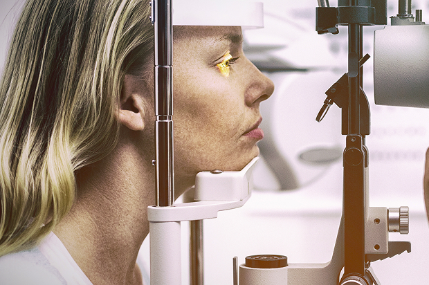
At "K+31," retinitis diagnosis begins with a conversation and an examination, but does not end there. The doctor checks vision, examines the fundus, and selects instrumental tests. The goal is to confirm retinal inflammation, assess its activity, and find the cause.
Ophthalmologist examination and vision test
The doctor determines when the floaters, flashes, fog, photophobia, or decreased visual acuity appeared. Then, the following is performed:
- Vision test
- Intraocular pressure measurement
- Examination of the anterior segment of the eye
These findings help determine whether there are signs of uveitis or other inflammation.

Fundus examination, OCT, and angiography
Diagnostics reveals foci of inflammation, hemorrhage, edema, and vascular changes. OCT evaluates the retina layer by layer and helps identify edema in the central area. Angiography is necessary to understand the functioning of the vascular network and whether there are areas of impaired blood flow.
Tests and consultations with related specialists
If the doctor suspects an infection or an autoimmune process, tests and consultations with related specialists may be necessary. This is not a formality. Without finding the cause, retinitis treatment may be incomplete, and the inflammation may return.
How is retinitis different from other eye diseases?
Redness, fogginess, and discomfort are common with various eye conditions. Comparisons can help understand the general logic, but a definitive diagnosis can only be made by a doctor after a comprehensive examination.
How is retinitis different from other eye diseases?
| Symptom | Retinitis | Conjunctivitis | Uveitis |
|---|---|---|---|
| What is affected primarily? | Retina | Conjunctiva | Choroid |
| Main complaint | Deteriorating vision, flashes, floaters | Redness, discomfort, discharge | Pain, photophobia, blurred vision |
| Is urgent diagnosis necessary? | Yes | Usually depends on the situation | Yes |
The table shows that retinitis most often affects visual function. Conjunctivitis is more often characterized by superficial inflammation, while uveitis is characterized by pain and photophobia. However, these conditions can coexist, so self-diagnosis is unreliable.
Retinitis and conjunctivitis
Conjunctivitis affects the mucous membrane of the eye. It is characterized by redness, discharge, and a gritty sensation. Retinitis is characterized by blurred vision, flashes, and loss of image areas.
Uveitis and retinitis
Uveitis is an inflammation of the choroid. It most often causes pain, photophobia, and redness around the cornea. Retinitis can coexist with uveitis, so the doctor evaluates the entire eye, not just one complaint.
Chorioretinitis and retinitis
Chorioretinitis means that inflammation has affected the retina and choroid. This condition requires particularly careful vision evaluation, as the lesion can affect tissue nutrition and the restoration of visual function.
General information
How is retinitis treated?
Treatment is selected after an in-person examination. There is no universal treatment plan: infection, autoimmune inflammation, and the consequences of trauma require different solutions. The doctor explains to the patient the goal of each stage of therapy and how the results will be assessed.
Treatment of the cause of inflammation
If an infection is confirmed, therapy is directed at the causative agent. If the cause is autoimmune, control of the underlying systemic disease is important. When inflammation is associated with trauma or a complication of another disease, the treatment plan is tailored to the patient's overall condition.
Drug therapy
Medications can be used to control:
- Inflammation
- Infection
- Sedation
- Associated changes
The ophthalmologist selects the appropriate medications and regimens after diagnosis. Self-administering drops, antibiotics, or anti-inflammatory medications is dangerous: it can confuse the situation and waste time.
Result monitoring and follow-up monitoring
Follow-up monitoring shows whether the lesion is shrinking, whether the tissue structure is recovering according to OCT, and whether there are any new complaints. In "K+31," the patient receives not only a prescription but also a clear monitoring schedule. The treatment plan is adjusted if necessary.
"If retinitis is suspected, we don't limit ourselves to a single examination. It's important to quickly find the cause of the inflammation and assess the condition of the retina to avoid wasting time and preserve vision. Therefore, we use a comprehensive diagnostic approach and a personalized care plan for each patient."
— ophthalmologist



Prevention of retinitis depends on the cause. It's important to treat infections, manage chronic conditions, keep up with checkups for autoimmune conditions, and consult an ophthalmologist if new visual symptoms occur. After an inflammation episode, monitoring is necessary even if your condition improves.
FAQ
Can retinitis be cured?
What's the danger of retinitis?
How can you tell the difference between retinitis and conjunctivitis?
When should you urgently see a doctor?

Clinical Guidelines of the Ministry of Health of the Russian Federation — https://cr.minzdrav.gov.ru/
Russian Medical Journal, Ophthalmology Section — https://www.rmj.ru/articles/oftalmologiya/
Ophthalmology Journal / Media Sphere — https://www.mediasphera.ru/journal/oftalmologiya
Eypress portal - https://eyepress.ru/
Magazine “Attending Physician” - https://www.lvrach.ru/
MSD Manual in Russian - https://www.msdmanuals.com/ru
Our doctors





















This award is given to clinics with the highest ratings according to user ratings, a large number of requests from this site, and in the absence of critical violations.

This award is given to clinics with the highest ratings according to user ratings. It means that the place is known, loved, and definitely worth visiting.

The ProDoctors portal collected 500 thousand reviews, compiled a rating of doctors based on them and awarded the best. We are proud that our doctors are among those awarded.
Make an appointment at a convenient time on the nearest date
Price
Other services

Appointment to the doctor
Reviews





















Our clinics


Application “Personal Account K+31”












What is retinitis and why is it dangerous?
This is an inflammatory process in the retina of the eye. In medical literature, the retina is also called the retina. It lines the back of the eye and is responsible for image clarity, detail perception, light, and movement.
When the retina is inflamed, the cells of this tissue are damaged by infection, an immune response, a vascular disorder, or toxic damage. The doctor evaluates not only the lesion itself but also its location: changes in the central zone can more quickly affect reading, driving, and facial recognition.
Why retinal inflammation shouldn't be ignored
The retina doesn't hurt like the skin or mucous membranes do. Therefore, retinitis sometimes develops unnoticed, often without a person noticing:
Photopsies are flashes before the eyes, while scotomas are areas of the visual field where the image disappears completely or partially.
The main danger is damage to the visual cells and blood vessels. If the inflammation remains active for a long time, edema, scarring, chorioretinitis, or choroidal involvement can develop. Therefore, it is important for an ophthalmologist to quickly determine the cause and begin treatment for the inflammation under control.