
Retinopathy (retinal damage): symptoms, diagnosis, and treatment
Retinopathy usually develops without pain. A person may see normally for a long time. However, changes in the blood vessels and tissues of the retina are already occurring. The first noticeable signs are spots before the eyes, flashes of light, fog, distorted lines, or decreased vision.
If you experience such complaints, it's important not to wait for your vision to "recover on its own." An ophthalmologist examination is necessary.

specialists

equipment

treatment






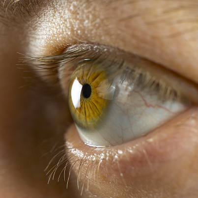
Types

Diabetic retinopathy
This type develops due to chronic vascular damage caused by diabetes. In the fundus, the doctor observes microaneurysms, macular edema, and areas of poor blood supply. Hemorrhages may also be present. Without monitoring, the condition can progress to severe vision loss.
Hypertensive retinopathy
Hypertensive retinopathy is associated with prolonged or severe hypertension. The retinal vessels narrow, their walls change, and edema sometimes occurs. An ophthalmological examination helps assess how hypertension affects the target organ.

Retinopathy of prematurity
Retinopathy of prematurity occurs in children whose retinal vessels have not yet fully formed. These patients require strict screening. The decision regarding observation, laser photocoagulation, or other treatment is made by a pediatric ophthalmologist based on the examination results.
Other forms of retinopathy
Retinal damage can occur after trauma, vascular accidents, radiation exposure, and severe general illnesses. The name of the form should always be based on the cause. Therefore, the diagnosis of retinopathy begins not with a predetermined treatment plan, but with identifying the source of the changes.
Symptoms you shouldn't ignore
Retinopathy symptoms can be mild, but their significance should not be underestimated. If the retina deteriorates gradually, the brain partially adapts to the deterioration in vision. The patient notices the problem only when the changes are already pronounced.

- Spots, flashes and fog before the eyes Floaters, flashes of light, and blurred vision are caused by changes in the retina, vitreous, or blood vessels. These symptoms do not point to a single diagnosis but require a fundus examination and OCT. It is especially important to seek medical attention promptly if the number of floaters increases sharply.
- Decreased visual acuity Vision loss associated with retinopathy may be due to central edema, hemorrhage, or tissue malnutrition. Sometimes, the patient has difficulty seeing small print, recognizing faces, and driving in dim light. The doctor assesses visual acuity and compares it with the examination data.
- Image distortion and visual field loss Image distortion, wavy lines, and visual field loss indicate involvement of important areas of the retina. In this situation, simply changing glasses is not enough. A retinopathy diagnosis with an assessment of the vessels and tissue structure is necessary.

How do we diagnose retinopathy?
At K+31, retinopathy diagnostics include an ophthalmologist examination and instrumental methods. Our goal is to visualize retinal changes, assess their activity, and understand the risk they pose to vision.
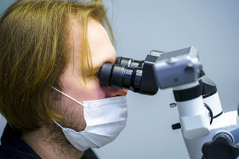
- Fundus examination A fundus examination reveals the condition of the vessels, the presence of hemorrhages, edema, and ischemic areas. The examination is performed after pupil dilation, unless contraindicated. The patient is explained the changes detected and why they are important.
- OCT of the retina OCT allows for a layer-by-layer assessment of the retina. This method is particularly useful when macular edema, thinning, or structural abnormalities are suspected. OCT does not replace a physical examination, but it does provide the physician with an accurate cross-sectional image.
- Fluorescein angiography Fluorescein angiography helps study blood flow and vascular permeability. The test is prescribed when it's necessary to pinpoint areas of ischemia, edema, or abnormal vascular growth. The decision is made by an ophthalmologist after an initial examination.
- Additional research Sometimes tonometry, ultrasound, laboratory tests, and consultations with related specialists are necessary. In diabetes, endocrinologist data and glycemic control are important, while in hypertension, blood pressure control is crucial. A team approach helps address the underlying cause, not just the ocular manifestations.
Modern treatment of retinopathy
Treatment for retinopathy is determined by its type, stage, and cause. The doctor assesses whether observation, laser photocoagulation, injection therapy, or surgery are needed. Sometimes, the key step is to stabilize the underlying condition.
Dynamic observation
With diabetic retinopathy, controlling blood sugar and associated risk factors is important. With hypertensive retinopathy, stabilizing blood pressure is essential. Without this, any topical treatment will be less effective because vascular damage will continue.
Laser coagulation
Laser coagulation is used when indicated to reduce the risk of progression and complications. This method is not suitable for everyone and does not automatically restore lost vision. Its purpose depends on the specific clinical situation.
Injection therapy
Injectable therapy, including anti-vascular growth factor agents, can be used for macular edema and certain vascular changes. The decision is made after diagnosis, taking into account the benefits, risks, and need for follow-up.
Surgical treatment
Vitrectomy is considered for severe complications, such as vitreous hemorrhage or traction changes. The extent of the procedure is determined on an individual basis. The patient receives an explanation in advance of the planned procedure and its purpose.
Types of retinopathy and how they differ
| Type | Cause | Main symptoms | Diagnosis | Treatment approach |
|---|---|---|---|---|
| Diabetic retinopathy | Diabetes mellitus | Floaters, decreased vision | OCT, fundus examination, angiography | Sugar control, laser, injections |
| Hypertensive retinopathy | Arterial hypertension | Decreased vision, retinal edema | Fundus examination, tonometry, OCT | Blood pressure monitoring, treatment of the underlying cause |
| Retinopathy of prematurity | Retinal vascular immaturity | Visual impairment, strabismus | Screening, fundus examination | Monitoring, laser coagulation as indicated |
"In our practice, we focus on early diagnosis: the sooner retinal changes are detected, the greater the chance of preserving vision. We conduct a comprehensive examination and tailor the treatment plan to your individual needs."
— ophthalmologist.
General information
Why patients choose us
Comprehensive diagnostics
We can assess the retina in a single visit: check vision, examine the fundus, perform an OCT scan, and determine the next step. This saves time and reduces uncertainty for the patient.
Experienced ophthalmologists
Our doctors treat various forms of retinopathy and understand when to involve an endocrinologist, a general practitioner, or a vitreoretinal surgeon. This approach is especially important for complex vascular changes.
Modern equipment
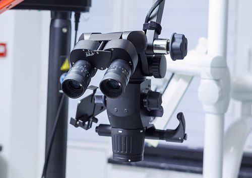
Instrumental diagnostics help us see what's impossible to assess based on complaints. We use the examination data for a personalized plan, not for formal diagnosis confirmation.
Personalized support
The patient receives a clear treatment plan, monitoring schedules, and an explanation of any alarming symptoms. If their condition changes, the treatment plan is reviewed.

FAQ
What is retinopathy in simple terms?
What symptoms of retinopathy should you be concerned about?
Is it possible to treat retinopathy without surgery?
How is retinopathy diagnosed?
Retinopathy requires attention. Even without pain, this condition can be painful. If you have diabetes, hypertension, vascular disorders, or new vision symptoms, schedule a consultation at K+31. An ophthalmologist will conduct an eye examination, explain the results, and suggest a plan for monitoring or treating your retinopathy.
Clinical Guidelines of the Ministry of Health of the Russian Federation — https://cr.minzdrav.gov.ru/
Ophthalmology Journal — https://www.mediasphera.ru/journal/oftalmologiya
Ophthalmology Bulletin Journal — https://www.mediasphera.ru/journal/vestnik-oftalmologii
Russian Medical Journal — https://www.rmj.ru/
Ophthalmology portal with specialized materials — https://eyepress.ru/
Attending Physician Journal — https://www.lvrach.ru/
Our doctors





















This award is given to clinics with the highest ratings according to user ratings, a large number of requests from this site, and in the absence of critical violations.

This award is given to clinics with the highest ratings according to user ratings. It means that the place is known, loved, and definitely worth visiting.

The ProDoctors portal collected 500 thousand reviews, compiled a rating of doctors based on them and awarded the best. We are proud that our doctors are among those awarded.
Make an appointment at a convenient time on the nearest date
Price
Other services

Appointment to the doctor
Reviews





















Our clinics


Application “Personal Account K+31”












Reasons
The primary mechanism is vascular damage and disruption of the retinal blood supply. In diabetes, the walls of small vessels become more vulnerable, leading to areas of edema and ischemia. In hypertension, vascular tone is impaired. Tissue nutrition may also be disrupted.
It is important for patients at risk to have their fundus examined regularly, even if their vision is not yet bothersome.