Retinal tear (retinal break): symptoms, diagnosis and treatment

A retinal tear is a condition in which a defect appears in the inner lining of the eye. If not treated promptly, fluid leaks under the lining. This leads to a detachment, leading to vision loss.

In our practice, we see that the earlier a retinal tear is detected, the higher the chance of a gentler treatment. In this article, we'll tell you how to recognize the problem, how diagnosis is performed, and why you shouldn't delay.

Best specialists
Best
specialists
Expert equipment
Expert
equipment
Advanced diagnostic treatment
Advanced diagnostic
treatment
Download the application “Personal Account K+31”
Everything about your health in one click!
Subscribe to social networks K+31
To keep up to date with current offers and receive useful advice about your health.

What is a retinal tear and why is it dangerous?

The retina is a thin, light-sensitive membrane that lines the inside of the eye and transmits images to the brain. When a defect develops, it means a retinal tear has formed. While small, it allows fluid to leak underneath the membrane.

The main danger is retinal detachment. Through the defect, fluid leaks under the membrane, lifting it. This causes rapid vision loss. Therefore, at the first warning signs, you should immediately consult a doctor while the process is still reversible.

What is a retinal tear and why is it dangerous?

Why does retinal detachment occur?

Most often, a retinal tear is associated with internal tension and peripheral thinning. Less commonly, it is caused by trauma. Let's look at the main mechanisms.

Myopia and thinning of the periphery of the shell

With myopia, the eye is elongated, and the peripheral membrane is stretched and thinned. These areas often become weak spots, where defects can more easily develop. Therefore, it is important for people with myopia to regularly examine the periphery of the fundus.

Eye trauma and physical activity

A direct blow, a concussion, or sudden physical exertion can damage the delicate membrane. Those with already weakened peripheral vision are especially at risk. After an eye injury, it's important to see a doctor. Don't delay the visit, even if your vision hasn't changed.

Age-related changes in the vitreous body

With age, the vitreous humor—the gel-like substance inside the eye—thickens and separates from the membrane. During this separation, the vitreous humor pulls on the membrane, which can cause a tear. This is one of the most common causes of this defect in older people.

How to recognize a retinal tear

How to recognize a retinal tear

The insidious thing is that the defect itself doesn't hurt. Characteristic visual symptoms of a retinal tear help to recognize it. The sooner you notice the symptoms of a retinal tear, the better your chances of preserving your vision.

Flashes, "lightning," and photopsia

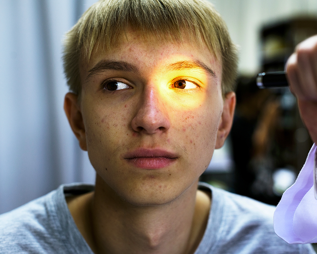
A common first sign is flashes before your eyes, especially noticeable in the dark or when moving your eyes. These are also called photopsies: they are short light signals caused by tension in the retinal membrane. When flashes appear before your eyes, the retinal membrane is experiencing traction, and this is a reason for an urgent examination. Persistent flashes before your eyes should not be ignored.
How to recognize a retinal tear

Floaters, floaters, and a "curtain" in front of the eye

The second typical sign is sudden floaters and spots in front of the eyes. A large number of these spots is called myodesopsia, and the sudden appearance of new floaters is alarming. If a shadow or "curtain" appears on the side of the visual field, this may indicate a detachment.

When is an urgent ophthalmologist consultation needed?

A warning sign is when flashes and floaters appear together and intensify. If a "curtain" or loss of part of the visual field is present, seek immediate medical attention. In such cases, an ophthalmologist will examine the eye the same day.

If you experience flashes, lightning, or sudden spots before your eyes, don't delay your appointment: the sooner a retinal tear is detected, the better your chance of preserving your vision.

General information

Diagnosing a retinal detachment

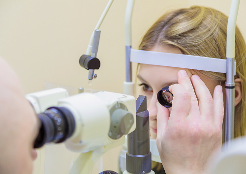
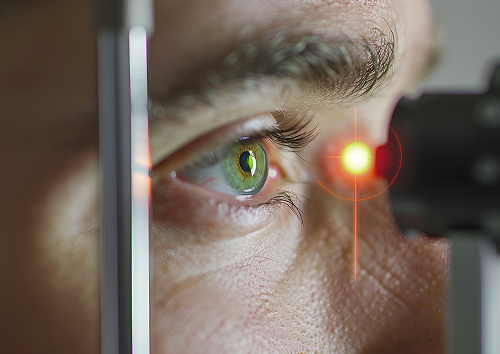
Diagnosing a retinal detachment begins with a detailed examination of the fundus. Our goal is to locate the retinal break, assess its location, and determine whether a retinal detachment has begun. The choice of treatment depends on this.

Fundus examination with pupil dilation

The key method is ophthalmoscopy with pupil dilation. After instilling drops, the pupil dilates, and the doctor examines the periphery of the retinal membrane, where the defect is most often hidden. Without pupil dilation, this examination is incomplete, so we perform it if a problem is suspected.

OCT, eye ultrasound, and other examination methods

For more information, OCT (optical coherence tomography) is used, which shows the retinal membrane layer by layer. If opacities interfere with the examination, an ocular ultrasound can be helpful. Together, OCT and ocular ultrasound provide a complete picture and identify hidden changes.

How a doctor assesses the risk of retinal detachment

The doctor checks for fluid around the defect and whether the vitreous is pulling on the retinal membrane. These signs indicate a high risk and require prompt attention. This is how we assess the likelihood of developing retinal detachment.
Diagnosing a retinal detachment

Methods for treating retinal detachments

Treatment for a retinal detachment depends on the location of the defect and whether detachment has already begun. If the process is detected early, gentler methods are often successful. We'll discuss these below, and include a table for clarity.

MethodWhen usedWhat benefits the patientRecovery features
Laser coagulation of the retinaFor a fresh tear without detachment"Welds" the defect, prevents detachmentOutpatient, fast, restrictions on physical activity for 2-3 weeks
CryopexyWhen laser is difficult, opacities, peripheryFreezing of tissue around the defect, creating a scarSwelling is possible, recovery takes up to 2-3 Weeks
Surgery (vitrectomy, buckling)For complications and detachmentRemoval of traction, repositioning of the retinaLong-term recovery, strict restrictions

Retinal laser coagulation

Retinal laser coagulation is the primary gentle method for treating a fresh defect without detachment. The laser creates microscars around the tear, which securely fix the retina and prevent fluid from penetrating underneath. The procedure is performed on an outpatient basis, is well-tolerated, and takes only a short time.

Cryopexy

Cryopexy is a cold treatment that freezes the tissue around the defect, creating a durable scar. This method is chosen when laser coagulation of the retina is difficult due to opacities or the specific location of the defect in the periphery. The result is similar to laser coagulation, but recovery can take up to two to three weeks.

Surgical treatment for complications

If a detachment has already developed, surgery is required: vitrectomy or buckling. The surgeon removes the tension and returns the membrane to its place. This treatment for a retinal tear is more complex, requires a long recovery period, and strict restrictions.

In our practice, we often see patients seeking help after flashes and a "curtain" appear before the eye. If a retinal tear is diagnosed early, it is often possible to stop the process using a gentle method and reduce the risk of detachment. This is why we emphasize rapid diagnosis and a clear treatment plan. – ophthalmologist

Recovery after cataract replacement

How we treat

We structure our treatment for retinal detachments so that the time from the first complaint to resolution is minimal. This is especially important when vision is at risk. We'll walk you through the process step by step.

Initial consultation and diagnosis

During the first appointment, the ophthalmologist clarifies the patient's complaints and performs a fundus examination with pupil dilation. If necessary, an OCT scan and ultrasound of the eye are prescribed. This diagnostic procedure quickly confirms the diagnosis and determines the location of the defect.

Choosing a treatment tactic

Based on the examination results, we select a treatment strategy: most often, laser coagulation, less commonly, cryopexy or surgery. The decision depends on whether there is a detachment and the location of the defect. We explain the treatment plan to the patient in understandable language.

Post-procedure monitoring

After the procedure, it's important to monitor the scar formation and the integrity of the membrane. We schedule follow-up appointments and provide recommendations on weight-bearing activities. This reduces the risk of recurrence.

How we treat

Our advantages

When vision is threatened, experience, equipment, and clear support are essential. We combine these in a single journey. Below is what the patient receives.

Experienced ophthalmologists

Our doctors regularly treat fundus pathology and quickly recognize dangerous changes in the periphery. This shortens the path to appropriate treatment. Experience helps us act decisively, even in urgent cases.

Modern equipment

Pupil dilation, OCT, and ultrasound are available during the appointment. All examinations are performed in one place and in a short time. This allows for rapid diagnosis confirmation.

Clear recommendations and patient support

We explain in simple terms what happened and what we do at each stage. The patient understands how to behave after the procedure and when to return for a follow-up. This kind of support reduces anxiety.

Our advantages

What to do before and after your doctor's appointment

Proper actions in the first few hours influence the outcome. Let's look at what you can and can't do before and after your examination.

What you can and can't do before your examination

Before your examination, avoid rubbing your eye, lifting heavy objects, or exercising. Don't self-diagnose or wait for the symptoms to go away on their own. If you experience flashes or spots, it's best to consult a doctor immediately.

Post-procedure restrictions

After laser or cryopexy, limit physical activity and heavy lifting for two to three weeks. Avoid bending over, saunas, and sudden movements. The doctor will determine the exact timing and rules individually.

When to return for a follow-up appointment

A follow-up appointment is necessary to ensure that the scar has formed and the membrane is stable. It's important to arrive on time. If flashes or spots reappear, you should contact us sooner.

What to do before and after your doctor's appointment

Who especially needs regular fundus examinations?

Some patients require routine fundus examinations more often than others. This helps detect dangerous changes early. Below are the patients who especially need urgent fundus examinations:

  • Patients with high myopia
  • Those who have had eye injuries
  • People with sudden flashes and floaters
  • Patients after ophthalmic surgery
  • Patients with peripheral retinal dystrophy

If you fall into these groups, it's important to have routine fundus examinations regularly.

Patients with nearsightedness

With nearsightedness, the periphery of the eye is thinned, and the risk of defects is higher. We recommend regular examinations with dilation for these patients. This is the basis for preventing retinal detachment.

After eye injuries and surgeries

A history of eye injury or surgery increases the risk of damage to the retinal detachment. Even with good vision, it's worth seeing a doctor for a checkup. This helps identify problems before they become complaints.

For peripheral retinal dystrophies

If peripheral retinal dystrophy has been previously diagnosed, areas of weakness require monitoring. Sometimes they are strengthened with laser prophylaxis. The doctor determines the appropriate course of action after an examination.

Who especially needs regular fundus examinations?

FAQ

Short answers to the questions most often asked during appointments.

What's the danger of a retinal tear?

This defect can lead to fluid leaking under the retinal membrane and retinal detachment. The sooner the patient seeks treatment, the higher the chance of stopping the process with a gentle approach. Therefore, there's no time to waste.

Why is a shadow on the side dangerous if vision in the center of the eye is still normal?

A shadow on the periphery may indicate the onset of a rhegmatogenous detachment. Fluid has already passed through the tear and is lifting the retina. As long as the central area is unaffected, the doctor has more options for preserving vision. Laser photocoagulation can sometimes help, but only under favorable conditions. If the detachment progresses further, treatment becomes more difficult, so complete vision loss should not be expected.

Is it possible to avoid surgery?

If the defect is detected early and detachment has not yet developed, laser coagulation or cryopexy are often used. The treatment strategy depends on the location of the defect and the condition of the membrane. The doctor will determine the exact plan.

What symptoms shouldn't be ignored?

Flashes before your eyes, lightning flashes, and sudden spots before your eyes, as well as loss of part of your visual field and a sensation of a "curtain," are dangerous. These are reasons to see an ophthalmologist immediately. These symptoms of a retinal tear require same-day examination.

What should you do after treatment?

Follow your doctor's instructions, avoid straining your eyes, and avoid strenuous physical activity. Be sure to return for a follow-up examination to confirm stable results. If new flare-ups or floaters appear, seek medical attention sooner.

List of sources

Clinical Guidelines Portal of the Ministry of Health of the Russian Federation — https://cr.minzdrav.gov.ru/

Federal Electronic Medical Library — https://femb.ru/

Ophthalmology Bulletin — https://www.mediasphera.ru/journal/vestnik-oftalmologii

Russian Medical Journal, Ophthalmology Section — https://www.rmj.ru/articles/oftalmologiya/

EyePress - https://eyepress.ru/

Our doctors

Karapetyan Arevik Samvelovna
Experience 12 years
Make an appointment
Karapetyan
Arevik Samvelovna
Ophthalmologist, Laser Surgeon
Batalina Larisa Vladimirovna
Experience 33 years
Make an appointment
Batalina
Larisa Vladimirovna
Deputy chief physician for ophthalmology, ophthalmic surgeon, leading specialist in laser vision correction
Amiryan Anush Gamletovna
Experience 28 years
Make an appointment
Amiryan
Anush Gamletovna
Ophthalmic oncologist surgeon
Medvedev Yulia Alexandrovna
Experience 11 years
Make an appointment
Medvedev
Yulia Alexandrovna
Ophthalmologist, Laser Surgeon
Rudkovskaya Elena Mikhailovna
Experience 38 years
Make an appointment
Rudkovskaya
Elena Mikhailovna
Ophthalmologist
Tegniryadnova Ekaterina Valerievna
Experience 20 years
Make an appointment
Tegniryadnova
Ekaterina Valerievna
Ophthalmologist, laser surgeon
Fadeeva Victoria Anatolievna
Experience 13 years
Make an appointment
Fadeeva
Victoria Anatolievna
Doctor ophthalmologist, laser surgeon
Shamsetdinova Leila Tagirovna
Experience 17 years
Make an appointment
Shamsetdinova
Leila Tagirovna
Pediatric ophthalmologist
Gavrilina Polina Dmitrievna
Experience 8 years
Make an appointment
Gavrilina
Polina Dmitrievna
Ophthalmologist, laser surgeon
Sugonyaeva Olga Yurievna
Experience 26 years
Make an appointment
Sugonyaeva
Olga Yurievna
Ophthalmologist, laser surgeon
Khlunovskaya Anna Nikolaevna
Experience 8 years
Make an appointment
Khlunovskaya
Anna Nikolaevna
Ophthalmologist
Akhiyarova Azalia Azatovna
Experience 4 years
Make an appointment
Akhiyarova
Azalia Azatovna
Ophthalmologist
Putintseva Polina Andreevna
Experience 6 years
Make an appointment
Putintseva
Polina Andreevna
Ophthalmologist
Agafonova Oksana Sergeevna
Experience 21 year
Make an appointment
Agafonova
Oksana Sergeevna
Ophthalmologist, pediatric
Kazennov Alexey Nikolaevich
Experience 19 years
Make an appointment
Kazennov
Alexey Nikolaevich
Ophthalmologist, ophthalmologist-surgeon
Petrova Maria Grigorievna
Experience 14 years
Make an appointment
Petrova
Maria Grigorievna
Ophthalmologist, ophthalmic surgeon
Mazmanyan Karen Akopovich
Experience 28 years
Make an appointment
Mazmanyan
Karen Akopovich
Ophthalmic surgeon
Suchkova Valeria Alekseevna
Experience 6 years
Make an appointment
Suchkova
Valeria Alekseevna
Ophthalmologist
Khubieva Salima Aslanbievna
Experience 7 years
Make an appointment
Khubieva
Salima Aslanbievna
Ophthalmologist, laser surgeon
Rytik Nina Petrovna
Experience 15 years
Make an appointment
Rytik
Nina Petrovna
Ophthalmologist
All specialists
2GIS Award
2GIS Award

This award is given to clinics with the highest ratings according to user ratings, a large number of requests from this site, and in the absence of critical violations.

«Good place» according to Yandex

This award is given to clinics with the highest ratings according to user ratings. It means that the place is known, loved, and definitely worth visiting.

Our doctors are laureates of the ProDoctors Award

The ProDoctors portal collected 500 thousand reviews, compiled a rating of doctors based on them and awarded the best. We are proud that our doctors are among those awarded.

Make an appointment at a convenient time on the nearest date

Price

Reception
Price
Initial Ophthalmologist Appointment
from 6 450 ₽
Corneal Collagen Crosslinking with Ultraviolet Irradiation
from 52 500 ₽
Femtosecond Laser-Assisted Intrastromal Segment Implantation
from 200 000 ₽

Other services

Laser vision correction Phacoemulsification of cataract with IOL implantation of any complexity Vitreoretinal surgeries of any complexity (vitrectomy) Laser photocoagulation of the retina during tears and degenerative changes in the retina Laser photocoagulation for diabetic retinopathy Intravitreal administration of Anti-VEGF drugs, hormonal implants Antiglaucomatous operations using various drains YAG laser dissection of the posterior capsule for secondary cataract Cataract treatment Treatment of glaucoma Retinal dystrophic changes Vitreous pathology Injury to the eye Post-traumatic and postoperative complications Myopia treatment Farsightedness Treatment of astigmatism Retinal disinsertion Non-contact laser procedure SMARTSURFACE Keratoplasty Femto SUPER LASIK Laser coagulation of the retina of the eye treatment on Ellex Integre Pro Scan Treatment of the retina with the Navilas laser system Femtolaser Cataract Removal Selection of children's night lenses Laser vision correction Clear Офтальмолог-онколог в Москве Treatment of keratoconus Laser coagulation of the retina Refractometry Retrobulbar injections Restoration of vision Lacrimal ducts Eye wash Synoptophore Forbis ESOM Pediatric ophthalmologist Ophthalmologist Glasses Biomicroscopy Night lenses Photo of an eye Eyelid suturing Selection of lenses Retinal laser Vision test Vitrectomy Vitrectomy Vitrectomy Eye pressure Ophthalmoscopy Fundus Gonioscopy Discision Orbitotomy Ophthalmodiagnostics OCT of the optic disc Keratoplasty Crystalline lens replacement Lacrimal duct Eye biometrics Angle of strabismus Lacrimal point IOL implantation Heterophoria Refraction Needling pillows Sclerectomy Micropulse laser Eyelid massage Intravitreal injections Iridoplasty Accommodation Bacterial culture Ophthalmology Cyclophotocoagulation Laser trabeculoplasty Laser stimulation Makdel Iridotomy Goniopuncture Corneal crosslinking Perimetry Pachymetry Angio-OCT Tubular test Color vision Convergence test Autofluorescence Eye mobility Corneal diagnostics Corneal transplant Skiascopy Strabismus Injection in the eye Tonometry Schirmer's test Trabeculotomy Diaphanoscopy Foreign body Foreign body Eyelid neoplasms Pterygium Chalazion Pneumatic retinopexy Glaucoma Ultrasound of the eye Eye biometrics Improving vision Cataract removal An attack of glaucoma Night lenses Eyelid ptosis Vision correction Angiography of the eye Retinoblastoma Laser coagulation
Make an appointment

Appointment to the doctor

Fill out the form, our managers will contact you within 15 minutes

Reviews

An excellent specialist. I am very pleased with the reception. Thanks to the doctor
08.07.2026
Svetlana U.
I would like to express my sincere gratitude to all the staff of the K+31 clinic. Special thanks to the doctors of the ophthalmology department, Ekaterina V. Tegniryadnova, who has been seeing me for a long time, and Maria Grigorievna Petrova, who operated on me for a lens replacement. Everything went great! I would also like to mention the work of Diana's ophthalmology manager. I received comprehensive and timely answers to all my questions. THANK you so much to all of you!
I am very pleased with the reception, smart doctor, thank you By +31.
26.06.2026
Irina T.
The Miracle Doctor
24.06.2026
Anush D.
I underwent preoperative preparation before laser correction at Azaliya Azatovna, I was satisfied! A very sensitive and understanding doctor. I drew schematically how the operation would take place, it was interesting and the fear went away. She also performed postoperative examinations. I wish you grateful patients and success in carrying out laser corrections. I will recommend it to my friends and family.
23.06.2026
Ekaterina Sh.
A very qualified doctor! Attentive and caring!
20.06.2026
Olga Ch.
A great doctor, explained everything, checked everything. I highly recommend a high-level specialist.
13.06.2026
Matvey S.
Почему К+31?
К + 31 — full-cycle multidisciplinary medical centers, including the possibility of providing medical services of European quality level.
К + 31 — are leading doctors and diagnostics using high-tech equipment from world manufacturers (Karl Storz, Olympus, Siemens, Toshiba, Bausch&Lomb, Technolas, Zeiss, Topcon).
К + 31 — is ethical. The staff of K+31 clinics maintain open relationships with patients and partners. An individual approach to each patient is the basis of our service standards.
К + 31 — is modernity. On call 24/7: call center operators will answer your questions at any time and book you an appointment with doctors. Contact us by phone, through the feedback form on the website and Max.

Our clinics

Address K+31 Lobachevskogo 2

st. Lobachevskogo, 42/7

Contacts

+7 499 999-31-31

Opening hours

Mon-Fri: 08:00 – 21:00
Weekend: 09:00 – 19:00

Address K+31 on Lobachevskogo

st. Lobachevskogo, 42/4

Contacts

+7 499 999-31-31

Opening hours

Mon-Fri: 08:00 – 21:00
Weekend: 09:00 – 19:00

Address K+31 Petrovskie Vorota

1st Kolobovsky pereulok, 4

Contacts

+7 499 999-31-31

Opening hours

Mon-Fri: 08:00 – 21:00
Sat-Sun: 09:00 – 19:00

Address K+31 West

Orshanskaya, 16/2; Ak. Pavlova, 22

Contacts

+7 499 999-31-31

Opening hours

Mon-Fri: 08:00 – 21:00
Sat-Sun: 09:00 – 18:00
K+31 on Lobachevskogo
Didn't find the service you were looking for?

Экстренная помощь