OCT of the Optic Nerve Head in Moscow

OCT of the optic nerve head is a modern diagnostic method that helps visualize the structure of the exit zone, assess the condition of nerve fibers, and detect changes that are not always visible during a routine examination. At our Moscow clinic, we perform the examination using modern equipment, and the results are evaluated by a doctor. Therefore, patients receive not only images and numbers but also a clear explanation of what they mean in their specific situation.

Best specialists
Best
specialists
Expert equipment
Expert
equipment
Advanced diagnostic treatment
Advanced diagnostic
treatment
Download the application “Personal Account K+31”
Everything about your health in one click!
Subscribe to social networks K+31
To keep up to date with current offers and receive useful advice about your health.

What is an optical examination of the optic nerve head?

Optical examination of the optic nerve head is performed using OCT. The full name of the method is optical coherence tomography. The device scans the eye tissue using a light beam and creates a layered image of the area being examined. The procedure requires no contact with the eye surface, is painless, and takes just a few minutes.

During the examination, the doctor obtains information about the appearance of the optic disc, the extent to which the nerve fiber layer is preserved, and whether there are any signs of thinning, swelling, or other structural changes. This approach is especially important for conditions where initial symptoms may not cause significant symptoms for a long time but can already impact the vision prognosis.

What is an optical examination of the optic nerve head?

When is OCT of the optic disc prescribed?

Indications for the test

The doctor may refer the patient for OCT of the optic disc if, during the examination, changes in the nerve exit zone are suspected or if monitoring of an existing condition is required.

When a doctor may refer for examination:

  • Suspected glaucoma or increased intraocular pressure
  • Vision deterioration without an obvious cause
  • Complaints of blurred vision, decreased clarity, or loss of image areas
  • Suspected disc edema
  • Signs of atrophy
  • Post-treatment follow-up
  • Hereditary predisposition to ophthalmological diseases

This examination of the optic nerve head is often required for more than just complaints. Sometimes the patient feels normal, but the ophthalmologist notices changes during the examination or is aware of risk factors, so they recommend further diagnostic testing.

Symptoms that require immediate diagnosis

Schedule an examination if you experience a narrowing of your visual field, blurred vision, difficulty finding your way in the dark, intermittent blurring, eye pain, a feeling of pressure, or frequent changes in glasses without consistent improvement.

Seek immediate medical attention if you experience a sudden decrease in vision, flashes of light, a cloudy vision, severe pain, severe redness, or a sudden deterioration in your overall condition.

How the study is conducted in our clinic

Preparing for OCT

The patient does not need to follow a diet, take any medications, or change their daily routine. If you wear contact lenses, remove them before the procedure to obtain a better image.

Sometimes, dilating drops are required for a complete examination, but this is not a requirement; the doctor will decide.

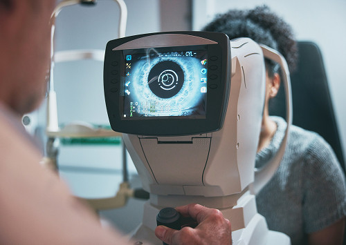
How the Procedure is Performed

All you need to do is fix your chin and forehead on the stand and look at the light mark. Try not to move or blink. During this time, the device performs the scan. There is no contact with the eye, so the procedure is painless and does not require anesthesia.

OCT of the optic disc is a quick procedure, but the quality of the result depends on proper gaze fixation, the transparency of the optical media, and the accuracy of the scan. If the image is insufficiently informative, a repeat scan is required to obtain data suitable for interpretation.

How the doctor interprets the results

After the examination, the doctor evaluates the images, numerical values, and color maps.

When interpreting the results, we consider the patient's age, eye structure, refraction, scan quality, examination data, and previous studies, if any.

General information

What does an OCT scan of the optic nerve head reveal?

Optic disc status

In the optic nerve head, retinal fibers gather and form the origin of the nerve that transmits visual information to the brain. Therefore, changes in this area can be an important diagnostic sign for various ophthalmological and neuro-ophthalmological conditions.

OCT of the optic nerve head helps assess the shape, size, and relationship of the retinal cup to the neuroretinal zonule, as well as other parameters that the doctor compares with the fundus examination. This information is especially important if glaucoma is suspected.

Retinal nerve fiber layer thickness

During this examination, the thickness of the retinal nerve fiber layer is assessed. These fibers are an extension of the cells that participate in signal transmission. In a number of diseases, they can become thinner, and the doctor's task is to detect such changes promptly.

The retina and optic nerve are functionally and anatomically connected, so when evaluating an image, it is important to look at the entire clinical picture, not just one section. If progression is suspected, we compare new data with previous results and evaluate the dynamics, not just a single value.

Signs of Glaucoma, Edema, and Atrophy

In glaucoma, attention is focused on the thinning of nerve fibers, changes in disc parameters, and the correspondence of these data to functional impairment. With disc edema, the picture is different: the increase in tissue volume, disc contours, and possible connection with other symptoms are assessed.

Atrophy also requires careful evaluation, as it can be caused by various reasons. In such cases, OCT helps to characterize structural changes, but further management may include additional methods and consultations with related specialists.

"In our practice, OCT of the optic nerve head is particularly valuable because it allows us to detect structural changes at an early stage. We recommend this examination not only for complaints but also for dynamic monitoring, when it's important not to miss even small changes."

What does an OCT scan of the optic nerve head reveal?

Why we recommend our examination

Modern diagnostic equipment

We use the latest equipment and can immediately obtain high-quality scans.

Optical coherence tomography is especially important for dynamic monitoring, because the doctor needs to compare indicators, see trends, and understand whether the situation is stable. The higher the quality of the scans, the more reliable the subsequent medical evaluation.

Accurate interpretation of results by an ophthalmologist

At our clinic, the results are evaluated by an ophthalmologist, who looks not only at the device printout but also at the patient. We explain which indicators are normal, which require monitoring, and which may be related to the structural features of the eye.

If the patient brings previous results, we compare them with the new data.

Convenient scheduling, quick examination, and clear recommendations

We schedule the procedure at a time that's convenient for you. The diagnostic process takes just a few minutes: you'll immediately receive an explanation of the results and recommendations.

At K31, you can undergo diagnostics in a single visit, if the clinical situation allows.

Why we recommend our examination

OCT, ophthalmoscopy, and perimetry: what's the difference?

What each method reveals

Ophthalmological methods don't compete with each other, but rather answer different questions. Ophthalmoscopy helps the doctor see the fundus and assess the appearance of the disc. Perimetry shows how the visual field is functioning. OCT provides layer-by-layer structural information and measurable parameters.

Method What it shows When it is prescribed Advantages
OCT of the optic nerve head Retinal disc structure and nerve fiber layer For suspected glaucoma, edema, or atrophy Highly informative, fast, and painless
Ophthalmoscopy Fundus appearance At the initial appointment An accessible basic examination
Perimetry Visual field To assess functional changes Helps monitor progress

Why examinations often complement each other

Structural changes may appear before functional ones, and sometimes complaints and visual field data require clarification using imaging. Therefore, OCT of the optic disc is often prescribed along with intraocular pressure measurement, fundus examination, and visual field testing.

OCT, ophthalmoscopy, and perimetry: what's the difference?

When should the examination be repeated?

Dynamic Monitoring in Glaucoma

If glaucoma is confirmed, the examination is repeated regularly because it is important for the doctor to determine whether the disease is stable or progressing.

Dynamic OCT of the optic disc allows for comparison with previous results. The doctor is interested not only in individual numbers, but also in the direction of changes, their speed, and their correlation with visual field data.

Monitoring after treatment or if progression of changes is suspected

Repeat examination of the optic nerve head may be necessary after a change in therapy, surgical or laser treatment, the appearance of new complaints, or a questionable result from a previous examination. Sometimes the doctor prescribes monitoring even in the absence of symptoms if risk factors are present.

When should the examination be repeated?

Frequently Asked Questions

What can be seen during the procedure?

OCT helps assess the condition of the optic disc, the thickness of the retinal nerve fiber layer, and identify structural changes that may be associated with glaucoma, edema, or atrophy. The final result is determined by the doctor after comparing it with the examination and complaints.

Do I need to prepare for the test?

Not usually, special preparation is required. Sometimes the doctor may recommend wearing contact lenses or temporarily removing them before the procedure to improve the quality of the scan.

Is OCT painful?

No, it is a contactless and painless test. The patient simply sits in front of the machine and looks at a marker while the machine performs the scan.

How often should I have an OCT of the optic nerve head?

The frequency depends on the diagnosis and the doctor's recommendations. For glaucoma and follow-up, the test may be scheduled regularly to monitor changes and promptly adjust treatment.
Sign up for an OCT scan of the optic nerve head in Moscow

Sign up for an OCT scan of the optic nerve head in Moscow

Schedule an appointment with K31 if you've been recommended an OCT scan of the optic disc, suspect optic nerve changes, have elevated intraocular pressure, a hereditary risk, or complain of vision deterioration. We'll conduct an examination using modern equipment, provide a medical interpretation, and explain next steps without complex language or unnecessary anxiety.

Our doctors

Batalina Larisa Vladimirovna
Experience 33 years
Make an appointment
Batalina
Larisa Vladimirovna
Deputy chief physician for ophthalmology, ophthalmic surgeon, leading specialist in laser vision correction
Amiryan Anush Gamletovna
Experience 28 years
Make an appointment
Amiryan
Anush Gamletovna
Ophthalmic oncologist surgeon
Medvedev Yulia Alexandrovna
Experience 11 years
Make an appointment
Medvedev
Yulia Alexandrovna
Ophthalmologist, Laser Surgeon
Rudkovskaya Elena Mikhailovna
Experience 38 years
Make an appointment
Rudkovskaya
Elena Mikhailovna
Ophthalmologist
Tegniryadnova Ekaterina Valerievna
Experience 20 years
Make an appointment
Tegniryadnova
Ekaterina Valerievna
Ophthalmologist, laser surgeon
Fadeeva Victoria Anatolievna
Experience 13 years
Make an appointment
Fadeeva
Victoria Anatolievna
Doctor ophthalmologist, laser surgeon
Shamsetdinova Leila Tagirovna
Experience 17 years
Make an appointment
Shamsetdinova
Leila Tagirovna
Pediatric ophthalmologist
Gavrilina Polina Dmitrievna
Experience 8 years
Make an appointment
Gavrilina
Polina Dmitrievna
Ophthalmologist, laser surgeon
Kovaleva Olesya Alexandrovna
Experience 7 years
Make an appointment
Kovaleva
Olesya Alexandrovna
Ophthalmologist, laser surgeon
Sugonyaeva Olga Yurievna
Experience 26 years
Make an appointment
Sugonyaeva
Olga Yurievna
Ophthalmologist, laser surgeon
Khlunovskaya Anna Nikolaevna
Experience 8 years
Make an appointment
Khlunovskaya
Anna Nikolaevna
Ophthalmologist
Akhiyarova Azalia Azatovna
Experience 4 years
Make an appointment
Akhiyarova
Azalia Azatovna
Ophthalmologist
Putintseva Polina Andreevna
Experience 6 years
Make an appointment
Putintseva
Polina Andreevna
Ophthalmologist
Agafonova Oksana Sergeevna
Experience 21 year
Make an appointment
Agafonova
Oksana Sergeevna
Ophthalmologist, pediatric
Kazennov Alexey Nikolaevich
Experience 19 years
Make an appointment
Kazennov
Alexey Nikolaevich
Ophthalmologist, ophthalmologist-surgeon
Petrova Maria Grigorievna
Experience 14 years
Make an appointment
Petrova
Maria Grigorievna
Ophthalmologist, ophthalmic surgeon
Mazmanyan Karen Akopovich
Experience 28 years
Make an appointment
Mazmanyan
Karen Akopovich
Ophthalmic surgeon
Suchkova Valeria Alekseevna
Experience 6 years
Make an appointment
Suchkova
Valeria Alekseevna
Ophthalmologist
Khubieva Salima Aslanbievna
Experience 7 years
Make an appointment
Khubieva
Salima Aslanbievna
Ophthalmologist, laser surgeon
Rytik Nina Petrovna
Experience 15 years
Make an appointment
Rytik
Nina Petrovna
Ophthalmologist
All specialists
2GIS Award
2GIS Award

This award is given to clinics with the highest ratings according to user ratings, a large number of requests from this site, and in the absence of critical violations.

«Good place» according to Yandex

This award is given to clinics with the highest ratings according to user ratings. It means that the place is known, loved, and definitely worth visiting.

Our doctors are laureates of the ProDoctors Award

The ProDoctors portal collected 500 thousand reviews, compiled a rating of doctors based on them and awarded the best. We are proud that our doctors are among those awarded.

Make an appointment at a convenient time on the nearest date

Price

Reception
Price
Initial Ophthalmologist Appointment
from 6 450 ₽
Corneal Collagen Crosslinking with Ultraviolet Irradiation
from 52 500 ₽
Femtosecond Laser-Assisted Intrastromal Segment Implantation
from 200 000 ₽

Other services

Laser vision correction Phacoemulsification of cataract with IOL implantation of any complexity Vitreoretinal surgeries of any complexity (vitrectomy) Laser photocoagulation of the retina during tears and degenerative changes in the retina Laser photocoagulation for diabetic retinopathy Intravitreal administration of Anti-VEGF drugs, hormonal implants Antiglaucomatous operations using various drains YAG laser dissection of the posterior capsule for secondary cataract Cataract treatment Treatment of glaucoma Retinal dystrophic changes Vitreous pathology Injury to the eye Post-traumatic and postoperative complications Myopia treatment Farsightedness Treatment of astigmatism Retinal disinsertion Non-contact laser procedure SMARTSURFACE Keratoplasty Femto SUPER LASIK Laser coagulation of the retina of the eye treatment on Ellex Integre Pro Scan Treatment of the retina with the Navilas laser system Femtolaser Cataract Removal Selection of children's night lenses Laser vision correction Clear Офтальмолог-онколог в Москве Treatment of keratoconus Laser coagulation of the retina Refractometry Retrobulbar injections Restoration of vision Lacrimal ducts Eye wash Synoptophore Forbis ESOM Pediatric ophthalmologist Ophthalmologist Glasses Biomicroscopy Night lenses Photo of an eye Eyelid suturing Selection of lenses Retinal laser Vision test Vitrectomy Vitrectomy Vitrectomy Eye pressure Ophthalmoscopy Fundus Gonioscopy Discision Orbitotomy Ophthalmodiagnostics Keratoplasty Crystalline lens replacement Lacrimal duct Eye biometrics Angle of strabismus Lacrimal point IOL implantation Heterophoria Refraction Needling pillows Sclerectomy Micropulse laser Eyelid massage Intravitreal injections Iridoplasty Accommodation Bacterial culture Ophthalmology Cyclophotocoagulation Laser trabeculoplasty Laser stimulation Makdel Iridotomy Goniopuncture Corneal crosslinking Perimetry Pachymetry Tubular test
Make an appointment

Appointment to the doctor

Fill out the form, our managers will contact you within 15 minutes

Reviews

The Miracle Doctor
24.06.2026
Anush D.
I underwent preoperative preparation before laser correction at Azaliya Azatovna, I was satisfied! A very sensitive and understanding doctor. I drew schematically how the operation would take place, it was interesting and the fear went away. She also performed postoperative examinations. I wish you grateful patients and success in carrying out laser corrections. I will recommend it to my friends and family.
23.06.2026
Ekaterina Sh.
A very qualified doctor! Attentive and caring!
20.06.2026
Olga Ch.
Olesya Alexandrovna is an excellent specialist, listened attentively and carried out the necessary diagnostics, answered all questions in detail and kindly and gave recommendations. I plan to see Dr. Kovaleva in the future.
18.06.2026
Anton Sh.
A great doctor, explained everything, checked everything. I highly recommend a high-level specialist.
13.06.2026
Matvey S.
Everything is fine, thanks again.
09.06.2026
Vladimir S.
A great doctor!!!
03.06.2026
Darya N.
We are patients of the clinic aged K+31, 89 and 85. The YUI clinic recommended cataract surgery. Of course, unrest and fears began. Everything started well and correctly with a call to Diana, the manager of the ophthalmology department. In a soft, calm voice, Diana explained everything clearly and provided real help and support when arranging medical services. Diana's attention and, one might say, her care have been very supportive. Diana made an appointment for YUI with a very good doctor Petrova Maria Grigoryevna. We are grateful to Diana for this attitude and her clear work!
01.06.2026
Yuriy B.
Tegniryadnova is an excellent doctor, we have done a lot of research, but the clinic is cold and uncomfortable
27.05.2026
Yu. Mir.
Excellent doctor Podshchekoldina E.N., Tegniryadnova E.V. the best ophthalmologist
Почему К+31?
К + 31 — full-cycle multidisciplinary medical centers, including the possibility of providing medical services of European quality level.
К + 31 — are leading doctors and diagnostics using high-tech equipment from world manufacturers (Karl Storz, Olympus, Siemens, Toshiba, Bausch&Lomb, Technolas, Zeiss, Topcon).
К + 31 — is ethical. The staff of K+31 clinics maintain open relationships with patients and partners. An individual approach to each patient is the basis of our service standards.
К + 31 — is modernity. On call 24/7: call center operators will answer your questions at any time and book you an appointment with doctors. Contact us by phone, through the feedback form on the website and Max.

Our clinics

Address K+31 Lobachevskogo 2

st. Lobachevskogo, 42/7

Contacts

+7 499 999-31-31

Opening hours

Mon-Fri: 08:00 – 21:00
Weekend: 09:00 – 19:00

Address K+31 on Lobachevskogo

st. Lobachevskogo, 42/4

Contacts

+7 499 999-31-31

Opening hours

Mon-Fri: 08:00 – 21:00
Weekend: 09:00 – 19:00

Address K+31 Petrovskie Vorota

1st Kolobovsky pereulok, 4

Contacts

+7 499 999-31-31

Opening hours

Mon-Fri: 08:00 – 21:00
Sat-Sun: 09:00 – 19:00

Address K+31 West

Orshanskaya, 16/2; Ak. Pavlova, 22

Contacts

+7 499 999-31-31

Opening hours

Mon-Fri: 08:00 – 21:00
Sat-Sun: 09:00 – 18:00
K+31 on Lobachevskogo
Didn't find the service you were looking for?

Экстренная помощь