
Focal Laser Photocoagulation of the Fundus and Retina in Moscow
When retinal vessels begin to leak fluid or localized risk areas appear on the retina, it is important for the doctor to accurately determine the location of the lesion and its potential for vision risk.
At K+31, focal laser photocoagulation of the fundus in Moscow is performed only after an in-person examination, comprehensive diagnostics, and an assessment of the macular area. We explain the purpose of the procedure to the patient in advance and do not promise the same results for everyone.
The goal of treatment is to stabilize the affected area, reduce the risk of progression, and help preserve vision as much as the eye's original condition allows.

specialists

equipment

treatment






In what cases do we recommend the procedure?
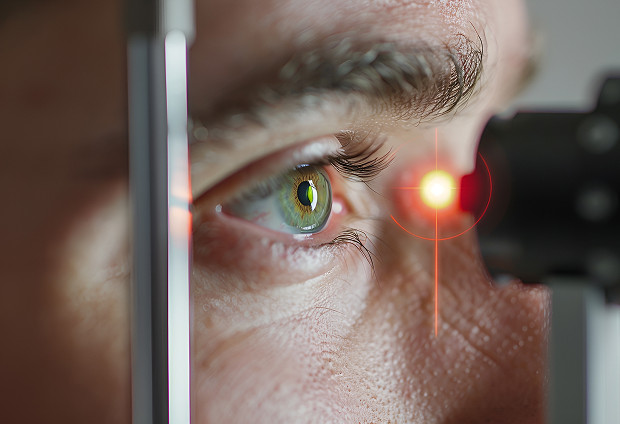
A laser isn't prescribed for every change. First, the doctor determines the location of the lesion, its activity, whether there's a threat to the central area, and the severity of vision impairment. Indications depend on:
- The cause of the disease
- The depth of the lesion
- The risk of progression
Diabetic Retinopathy and Macular Edema
Diabetes can damage small blood vessels in the eye. This leads to diabetic retinopathy, and fluid sometimes accumulates in the macula. If macular edema develops, a person may notice blurring and decreased clarity in the center of the visual field.
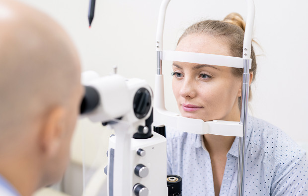
A focal laser is considered when the leakage zone is accessible for precise treatment. However, diabetic retinopathy requires monitoring over time. If the edema affects the macula, the treatment strategy depends on its location, severity, and impact on the central part of the eye.
Local retinal changes and other indications
The procedure may be recommended for limited vascular changes, peripheral lesions, or areas where the retina requires preventative strengthening. The doctor evaluates the lesion, adjacent tissues, and the transparency of the optical media.
If the risk is low, we choose observation. If the indications are confirmed, we explain the purpose of the laser and the limitations of the method.
How focal laser coagulation of the fundus is performed at our center
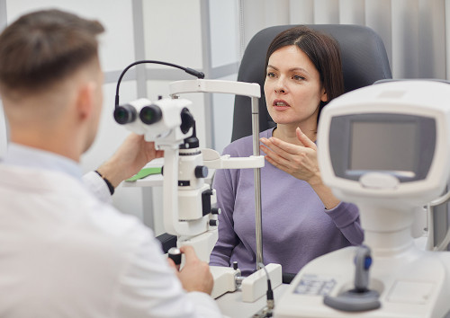
Before laser photocoagulation, we clarify the patient's complaints, illness duration, chronic diagnoses, medications, and allergies. The doctor then assesses the retina's appearance with the pupil dilated, including any swelling, hemorrhages, vascular changes, or areas of thinning. Without this step, precise treatment is impossible.
Pre-procedure diagnostics
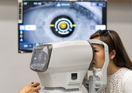
Vision diagnostics are necessary to avoid a "rough" treatment. We check visual acuity and intraocular pressure, conduct a pupillary dilation examination, and, if necessary, order a tomography scan. This allows the doctor to see the location of the lesion and the fundus.
Preparation includes steps that help confirm the diagnosis and assess safety:
- Ophthalmologist consultation
- Retina diagnostics
- Checking indications and contraindications
- Laser parameter selection
- Post-procedure recommendations
After preparation, the patient understands the procedure. If there is insufficient information, treatment is postponed until the condition is clarified.
Laser Treatment Stages
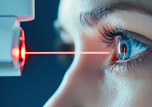
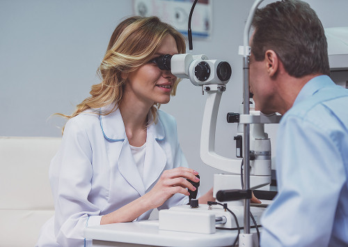
The session is performed at an outpatient clinic. Pupil dilation and local anesthesia are administered, then the doctor inserts a lens to view the treatment area. The laser then delivers short pulses to the selected points.
We consider laser retinal treatment as part of an individualized plan, not a one-size-fits-all procedure. Parameters depend on the location of the lesion, the severity of the swelling, and the transparency of the media. If the retinal changes are near the macula, precision is especially important.
What does the patient experience during the session?
The procedure is usually well-tolerated. The patient sees flashes of light, feels the lens touch, and sometimes feels brief pressure or mild discomfort. Severe pain is not typical for this method. After the session, the following may occur:
- Light sensitivity
- Tearing
- Temporary blurring
- A feeling of eye fatigue
These reactions are most often associated with pupil dilation and local stimulation. If laser retinal coagulation is prescribed, the doctor will explain the expected sensations in advance.
General information
Advantages of Focal Laser Coagulation
The main advantage of this method is its localization. The doctor treats a specific area, rather than a large, unnecessarily large area. When retinal changes are focal, this approach allows for more precise treatment.
Targeted action on the pathological area
The laser is directed to the location where there is a medical reason for treatment: an area of fluid leakage, a vascular defect, or an area supporting edema. With vascular diseases, precision is especially important because the changes are often unevenly distributed.
Doctor's quote:
"In our experience, the key to a successful outcome is an accurate diagnosis before the procedure." We tailor laser parameters individually to target only the desired area and preserve healthy retinal tissue."
Preservation of Healthy Retinal Tissue
The focal principle reduces unnecessary exposure to adjacent areas. This is important for the patient because the retina is involved in perceiving light, contrast, shape, and detail. The more precisely the area is selected, the more accurately the treatment is performed.
The laser does not treat the underlying disease. If the vascular process remains active, monitoring of the results is necessary, and sometimes medications or other interventions are needed. The doctor explains when laser treatment of the retina is aimed at stabilization.
Outpatient format and short recovery
The procedure is usually performed on an outpatient basis, without hospitalization. The patient receives recommendations, a follow-up date, and restrictions. The early period often proceeds without prolonged disruption to the usual routine, but the timing varies from person to person.
Comparison helps understand why the doctor chooses focal treatment. The final decision is still made after diagnosis.
| Method | When used | What it provides | After treatment |
|---|---|---|---|
| Focal photocoagulation | Local lesion | Stabilization of the risk zone | Usually a short period |
| Observation | Low risk | Monitoring without intervention | No special Restrictions |
| Medications | Edema, vascular activity | Edema reduction | Repeat visits are required |
| Panretinal coagulation | Common process | Complication prevention | May take longer |
The table does not replace a consultation. For some patients, observation is sufficient, for others, a different laser technique is needed, and for others, a combined plan is needed.
Frequently Asked Questions
Before the procedure, ophthalmology patients most often ask about pain, timing, and results. It's best to get answers in advance.
Is the procedure painful?
How long does the post-procedure period last?
When are results visible?
Are there any restrictions to the procedure?
Sign up for a consultation

If the examination reveals vascular changes, edema, or a localized risk to the retina, you should not choose treatment on your own. Focal laser coagulation of the fundus is only useful with a confirmed diagnosis, a precise target area, and a clear goal.
At K+31, the doctor will compare complaints and images, evaluate the fundus, and suggest a safe route.
During the appointment, we check visual acuity, evaluate the retina in different areas, and discuss previous findings. If laser coagulation of the retina is necessary, the patient understands the procedure and post-treatment guidelines in advance.
Our doctors




















This award is given to clinics with the highest ratings according to user ratings, a large number of requests from this site, and in the absence of critical violations.

This award is given to clinics with the highest ratings according to user ratings. It means that the place is known, loved, and definitely worth visiting.

The ProDoctors portal collected 500 thousand reviews, compiled a rating of doctors based on them and awarded the best. We are proud that our doctors are among those awarded.
Make an appointment at a convenient time on the nearest date
Price
Other services

Appointment to the doctor
Reviews





















Our clinics


Application “Personal Account K+31”












What is focal laser coagulation and why is it needed?
Focal coagulation is a targeted laser treatment of a limited area. The doctor determines the treatment points in advance, assessing the vessels, macula, and fundus. Essentially, laser coagulation of the retina helps treat:
The term focal photocoagulation is found in medical documents. The meaning is the same: the laser targets specific points. The decision is made by an ophthalmologist after an examination, as the retina does not always produce obvious symptoms early on.