Eye biomicroscopy in Moscow – accurate slit lamp diagnostics
Timely vision diagnostics allow us to detect many diseases at an early stage. Our clinic offers a detailed slit lamp examination, which provides the doctor with a complete clinical picture of the optical environment.
We perform this examination daily, helping patients maintain their health and quality of life. Modern eye biomicroscopy is available to residents and visitors of Moscow.

specialists

equipment

treatment


When is biomicroscopy prescribed?
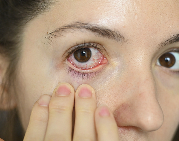
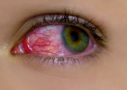
Patients often seek our help for vision diagnostics if they experience redness, severe burning, or severe photophobia, as well as a gritty sensation under the eyelids. In these situations, biomicroscopy of the eye helps quickly identify the underlying cause of the discomfort.
Biomicroscopy after injuries and surgeries
Any mechanical or chemical eye injury requires immediate instrumental monitoring of the tissue condition. We carefully assess the extent of damage to determine the appropriate patient management strategy. After surgery, regular monitoring helps doctors monitor the natural healing process.
Monitoring chronic eye diseases
Patients with chronic pathologies require ongoing professional monitoring. Regular eye examinations allow us to promptly detect hidden exacerbations of the condition. This enables us to promptly adjust therapy before serious structural complications develop.
How is eye biomicroscopy performed?
Typically, no complicated preparation is required; simply arrive at the clinic on time. If you wear contact lenses, we will ask you to remove them before the slit lamp examination. Sometimes, to better visualize internal areas, we administer pupil dilation drops. It's important to note that after using these medications, overall visual acuity may be temporarily reduced.
Stages of the procedure
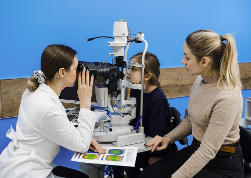
The patient sits in front of the device and rests their chin on a special ergonomic rest. The ophthalmologist then adjusts comfortable lighting and begins the examination through the microscope. We smoothly move the light beam, sequentially examining all necessary tissue areas.
How long does the examination take?
The diagnostic process itself takes literally a few minutes and is completely stress-free. If the pupil requires dilation, you'll have to wait about fifteen minutes in the hallway. In any case, we strive to complete the procedure as quickly and efficiently as possible.
What does the patient feel during the examination?
The procedure is as comfortable as possible; the patient only feels a bright light directed at their face. We always warn them that blinking is allowed and recommended to avoid overdrying the mucous membrane. There are no painful or unpleasant physical effects during the examination.
General information
What can biomicroscopy reveal?
Serious eye inflammation requiring medical treatment often lurks behind the guise of simple fatigue. With a focused examination, we easily diagnose conditions such as keratitis or chronic blepharitis.
If the deep vascular layers are affected, the device will immediately reveal characteristic signs indicating uveitis.
Corneal and conjunctival damage
The superficial layers are the first to suffer from adverse external factors and dehydration. We carefully examine the cornea for microscopic erosions, ulcers, or degenerative changes.
We also carefully evaluate the conjunctiva to completely rule out hidden allergic reactions and microtraumas.
Changes in the lens and signs of cataracts
With age, a person's natural lens gradually begins to lose its original transparency. Early detection of cataracts allows for the correct planning of surgery at the optimal time. We examine the lens under different illumination angles, accurately determining the degree and location of opacities.
Foreign bodies and consequences of trauma
Even the tiniest foreign body can cause severe pain and profuse lacrimation. Under high magnification, we accurately determine the depth of its penetration into the tissue. This ensures completely safe and complete removal of foreign particles without the risk of complications.
FAQ
Is an eye biomicroscopy painful?
Do I need to prepare for a biomicroscopy?
What does eye biomicroscopy reveal?
How long does the examination take?

We have combined cutting-edge diagnostic equipment and the highly professionalism of our doctors in one location. Our friendly team is truly committed to the psychological well-being of every clinic visitor. We guarantee that you will receive honest, comprehensive, and understandable information about your health.
How to make an appointment
To choose the most convenient time, simply call our reception desk or submit a simple request on our website. Our friendly receptionists will answer all your organizational questions and help you schedule an appointment with the right specialist.
Remember, regular preventive examinations are the best investment in your vision.
Clinical Guidelines of the Ministry of Health of the Russian Federation — https://cr.minzdrav.gov.ru/
Russian Society of Ophthalmologists — https://roou.ru/
MSD Manuals in Russian — https://www.msdmanuals.com/ru
CyberLeninka — Publications on Ocular Biomicroscopy — link
CyberLeninka - materials on the slit lamp in ophthalmology - link
eLIBRARY - scientific publications on the topic - link
Our doctors


This award is given to clinics with the highest ratings according to user ratings, a large number of requests from this site, and in the absence of critical violations.

This award is given to clinics with the highest ratings according to user ratings. It means that the place is known, loved, and definitely worth visiting.

The ProDoctors portal collected 500 thousand reviews, compiled a rating of doctors based on them and awarded the best. We are proud that our doctors are among those awarded.
Make an appointment at a convenient time on the nearest date
Price
Other services

Appointment to the doctor
Reviews


Our clinics

Application “Personal Account K+31”


What structures does the doctor examine?
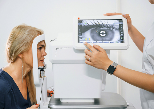
To make an accurate diagnosis, it is necessary to examine the anterior segment of the eye in detail. Using a focused light beam and a binocular microscope, we obtain contrast optical magnification. Before listing the inspection areas, it is important to note that this method allows for a layer-by-layer examination of tissue.
Here are the main elements the doctor evaluates during the procedure:
Such a detailed eye examination eliminates accidental errors during the initial assessment of structures. Every detail examined is crucial for making an accurate diagnosis.
How does biomicroscopy differ from a routine examination?
A standard ophthalmological examination only provides a general picture of the visual system. Detailed biomicroscopy allows for the examination of minimal tissue changes that are hidden from the naked eye.