
Barrier Laser Photocoagulation of the Retina
The retina is responsible for image perception and the transmission of visual information to the brain. If areas of thinning or damage appear in its structure, the risk of serious complications increases. Barrier laser photocoagulation of the retina is considered one method for preventing such conditions. The procedure helps strengthen weak areas and reduce the risk of further progression of changes.
In our practice, barrier laser photocoagulation is performed only after a complete examination. First, we assess the overall condition, determine the degree of risk, and only then decide on the need for treatment.

specialists

equipment

treatment






When a doctor recommends a procedure

A prescription is only possible after an examination and assessment of the fundus. Myopia or age-related changes alone are not grounds for treatment.
Peripheral Retinal Dystrophy
Peripheral retinal dystrophy is considered one of the most common reasons for prescription.
The danger is that a person may not notice any symptoms for a long time. Changes are only detected during an examination, when the doctor examines the fundus after dilating the pupil.
Tears, Thinning, and Other Risks
Retinal tears, localized areas of degeneration, and retinal thinning may be indications for treatment. Such changes increase the risk of serious complications, especially with physical activity or disease progression.
If a small retinal tear is detected during an examination, timely laser coagulation can often avoid more complex treatment later.

Prevention of Retinal Detachment
The primary goal of the procedure is to prevent retinal detachment. This complication is considered one of the most dangerous for vision.
If the doctor identifies risk areas, timely tissue strengthening helps reduce the likelihood of further spread of the pathological process. It's important to understand that even a high-quality procedure does not provide an absolute guarantee.
The procedure may be considered in the following cases:
- Peripheral retinal dystrophy
- Retinal tears and thinning
- In case of increased risk of retinal detachment
- Before increased visual or physical stress
- After an examination, if the doctor detects dangerous changes
How is barrier laser coagulation performed?
An examination is mandatory before the procedure. Our goal is not only to confirm the diagnosis but also to assess the safety of future treatment.
Pre-procedure diagnostics
Preparation begins with a consultation with a doctor. The ophthalmologist assesses the patient's complaints, clarifies the medical history, and conducts an examination.
A mandatory part of the preparation is a fundus examination. For this, special drops are used to dilate the pupil and allow for a detailed examination of the peripheral areas.
If necessary, additional vision diagnostics are performed to help determine the scope of future treatment.
"In my experience, barrier laser coagulation is especially valuable when we identify risk areas early. This allows for targeted strengthening of the retina, without incisions, and with a rapid return to normal activity. For the patient, this is often the best way to avoid more serious complications."
Laser Treatment Stages
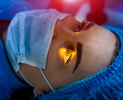
Before the procedure, the patient receives anesthetic drops. The doctor then inserts a special lens to help control the treatment area.
Laser photocoagulation is then performed on the affected areas. The laser creates a series of pinpoint coagulates around the risk zone. Gradually, a strong tissue bond is formed, limiting the spread of potential damage.
Modern laser photocoagulation is highly precise and allows for even minor adjustments.
What does the patient experience during the session?
The procedure is performed on an outpatient basis and is generally well-tolerated. Most patients report bright flashes of light and moderate discomfort during individual pulses.
There is generally no significant pain. Thanks to local anesthesia, the treatment is painless or accompanied by minimal discomfort.
General information
What happens after barrier coagulation?
After the procedure, the patient remains under the doctor's observation for a period of time and can then go home. Hospitalization is not required in most cases, as the treatment is performed on an outpatient basis.
In the first few hours, temporary light sensitivity, lacrimation, and visual discomfort are possible. These symptoms are considered expected and usually resolve on their own.
Restrictions and recommendations after the procedure
It takes time for strong coagulates to form. That's why it's important to follow your doctor's recommendations during the first few weeks.
We typically recommend:
- Limit strenuous physical activity
- Avoid heavy lifting
- Follow the prescribed monitoring regimen
- Keep follow-up appointments
- Closely monitor your well-being
Following recommendations helps reduce the risk of complications and ensure complete healing of the treated areas.
When is a follow-up appointment necessary?
The first follow-up appointment is scheduled at a time determined by your doctor. During this appointment, the condition and quality of the formed coagulates are assessed.
Another follow-up appointment may be necessary later, especially if the patient continues to have risk factors or significant changes in the periphery have been previously identified.




What to choose: observation, laser coagulation, or surgical treatment
| Method | When is it suitable? | Advantages | Limitations |
|---|---|---|---|
| Observation | If the risk of complications is minimal | Does not require intervention | Does not strengthen weak areas |
| Barrier Laser Coagulation | For dystrophies, ruptures, and risk areas | Pinpoint impact, minimally invasive, rapid recovery | Performed only when indicated |
| Surgical Treatment | For severe complications and established pathology | Allows for complex cases to be resolved | Longer recovery time after the procedure |
Frequently Asked Questions
Is barrier laser retinal coagulation painful?
How long does the procedure take?
Is any preparation necessary before barrier laser retinal coagulation?
What are the benefits of barrier laser retinal coagulation?
Conclusion: When you shouldn’t postpone a consultation

Diseases often develop without obvious symptoms. A person may not be aware of changes until serious complications arise.
If an examination reveals areas of thinning, signs of dystrophy, or peripheral changes, it is important to follow your doctor's recommendations and not delay treatment. Timely barrier laser coagulation of the retina helps strengthen tissue and reduce the likelihood of dangerous complications.
If you are bothered by flashes of light, new floaters have appeared, or changes in the fundus have been previously detected, schedule a consultation. Our ophthalmologist will conduct an examination, assess your condition, and determine the optimal monitoring or treatment plan.
Our doctors





















This award is given to clinics with the highest ratings according to user ratings, a large number of requests from this site, and in the absence of critical violations.

This award is given to clinics with the highest ratings according to user ratings. It means that the place is known, loved, and definitely worth visiting.

The ProDoctors portal collected 500 thousand reviews, compiled a rating of doctors based on them and awarded the best. We are proud that our doctors are among those awarded.
Make an appointment at a convenient time on the nearest date
Price
Other services

Appointment to the doctor
Reviews





















Our clinics


Application “Personal Account K+31”












Preparing for the procedure
In most cases, no special preparation is required. However, it is important to undergo a full examination and follow your doctor's recommendations before treatment.
We recommend checking in advance whether you will need assistance after the procedure. After pupil dilation, vision may remain blurred for some time, so driving is not recommended on the day of your procedure.
It is also important to inform your doctor about any medications you are taking and any chronic illnesses you have before treatment.