
Leukoma (eyesore): causes, symptoms and treatment
At the K+31 Clinic, we help patients with the most complex anterior segment pathologies every day. Modern eye microsurgery and innovative therapeutic approaches used by our specialists not only halt the pathological process but, in many cases, restore normal vision.

specialists

equipment

treatment






What is leukoma and why is it called an eyesore?
The human visual system is remarkably delicate, and its main optical element is the cornea—the transparent anterior layer of the eye. Normally, it perfectly transmits and refracts light rays, allowing us to see the world clearly and in detail. However, under the influence of various negative factors, this unique tissue can lose its properties, being replaced by opaque connective tissue fibers. In ophthalmology, such a persistent, pronounced change in corneal structure is called leukoma. This condition has long been known as an eyesore.
How corneal opacity forms
To understand how leukoma develops, it's important to understand the structure of the cornea. It consists of several layers, the most important of which is the stroma.
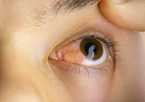
The stroma is formed by strictly oriented collagen sheets, which ensures the cornea's perfect transparency. If the tissue is deeply damaged—due to infection, mechanical stress, or exposure to harsh chemicals—the regeneration process is triggered. Unfortunately, the body is not always able to recreate its complex original structure. A dense, chaotically distributed corneal scar forms at the site of the damaged cells. Light can no longer pass freely through this area; it is scattered or completely blocked, causing a white spot to form on the surface of the eye.
How does leukoma differ from other eye diseases?
Not every corneal opacity can be immediately classified as a leukoma. Ophthalmologists classify opacities by their density, depth, and area of involvement. The mildest and most superficial change is called a cloud, while a more pronounced and noticeable change during a routine examination is called a macule.
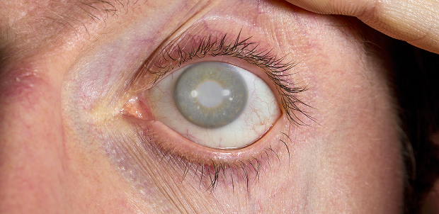
A leukoma, on the other hand, represents the final, densest, and most extensive stage of scarring, when fibrous tissue invades the deep layers of the stroma. Some patients mistakenly confuse it with a cataract. However, a cataract is a clouding of the crystalline lens, located inside the eye behind the pupil, while a corneal opacity is located on the very surface of the eye and is accessible to direct visual observation.
Why does leukoma appear?
After eye injuries and burns
One of the leading causes of scarring is mechanical trauma to the eye. This can include deep cuts, penetrating wounds, or exposure to scale or shavings when working with metal or wood without protective eyewear. Even more damaging is an eye burn, especially a chemical burn. Alkalis and acids instantly cause deep tissue necrosis, destroying the corneal epithelium and the limbal stem cells responsible for renewing the transparent layer.
Against a background of keratitis and corneal ulcers
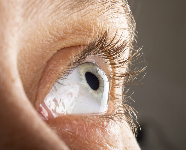
This condition is often triggered by advanced keratitis—an inflammation of the cornea caused by bacteria, viruses (especially herpes), or fungi. If the infection progresses, a corneal ulcer develops. This is a destructive condition in which the corneal tissue literally melts. After the acute inflammation subsides, the defect inevitably fills with coarse connective tissue, resulting in the formation of a dense cataract.
After inflammation, surgery, and tissue scarring
In some cases, the condition can develop after previous surgical interventions on the anterior segment of the eye, if healing was complicated or the patient has a congenital predisposition to excessive scarring. There are also congenital forms of the disease caused by intrauterine inflammatory processes or genetic developmental abnormalities.
What are the dangers of leukoma if left untreated?
Leukoma without regular monitoring by a specialist can become a source of serious medical problems.
Risk of persistent vision loss
If the scar is centrally located, prolonged lack of proper light stimulation of the retina can lead to the development of amblyopia (lazy eye syndrome), in which the brain simply loses the ability to perceive information from the affected visual organ.
Possible complications and scar progression
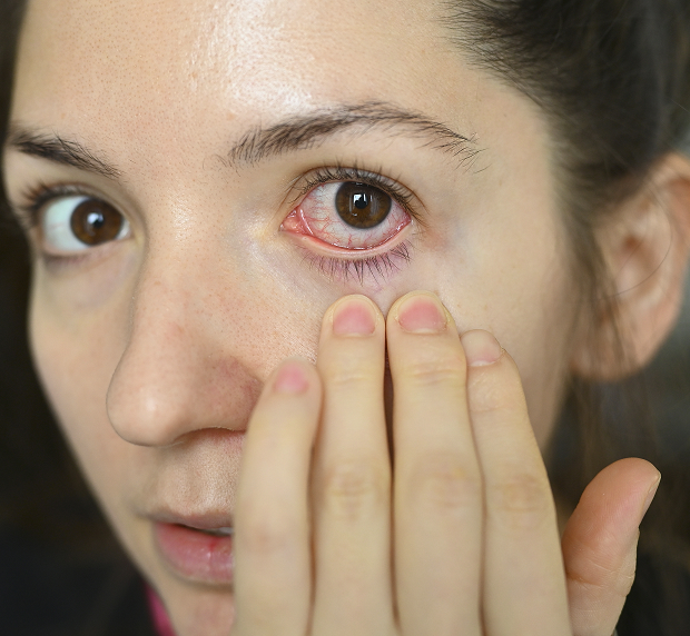
Dense scar tissue lacks normal blood supply and proper innervation. This makes it extremely vulnerable to any external microtrauma. Secondary erosions and trophic ulcers, which are prone to infection, can easily develop on the surface of the leukoma. In addition, newly formed vessels (neovascularization) often begin to grow into the scar tissue, which makes the scar even denser, contributes to its increase in size, and can cause repeated inflammatory attacks.
How a doctor diagnoses an eye spot
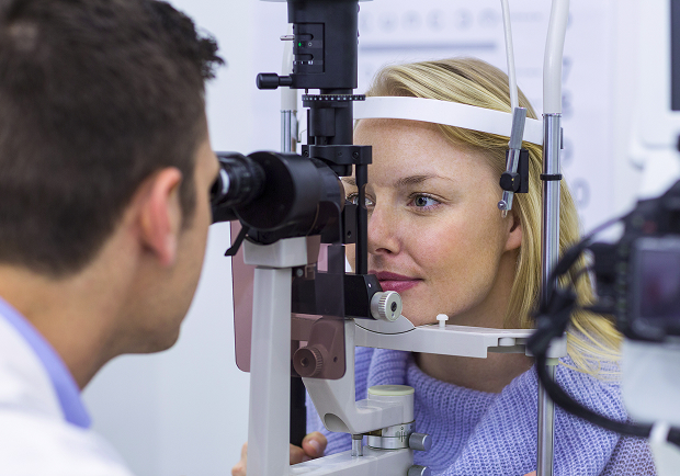
Treatment of leukoma is impossible without a comprehensive and in-depth examination. Comprehensive corneal diagnostics at the K+31 Clinic allow us to obtain comprehensive information about the patient's visual condition and develop the most effective and safe treatment plan.
Slit lamp examination
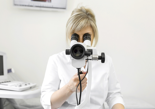
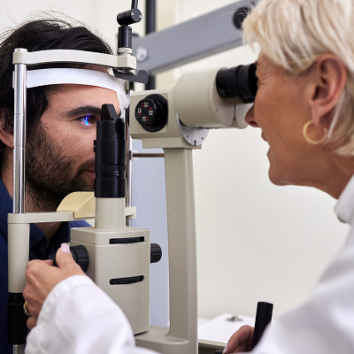
The most important and indispensable tool in our ophthalmologist's arsenal is the slit lamp. This instrument allows for high-magnification biomicroscopy of the eye using a special focused light beam. During this examination, the doctor can thoroughly examine the structure of the opacity, determine its precise boundaries, and assess how deeply the scar has penetrated the stromal layers.
Visual acuity and corneal health check
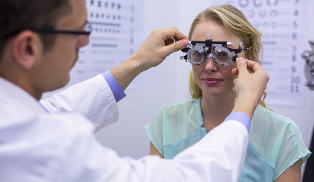
To assess vision and plan interventions, we perform visometry, corneal topography to create a three-dimensional map of the cornea, and pachymetry to measure corneal thickness.
Additional examination methods
If necessary, we supplement the examination with anterior segment OCT to accurately determine the depth of corneal scarring. We also use B-scan ultrasound to assess the condition of the lens, retina, and optic nerve for opacities.
Leukoma treatment methods
When conservative therapy helps
Conservative therapy with modern anti-inflammatory, regenerative, and enzymatic medications is indispensable during the development of opacity. This drug therapy helps to soften the maturing scar, although it cannot completely resolve long-standing changes.
Laser and microsurgical methods
For superficial opacities, the minimally invasive laser technique known as PTK is used to restore corneal transparency in a single day. The excimer laser gently vaporizes the affected tissue, ensuring rapid recovery.
Keratoplasty as a method for restoring corneal transparency
For deep, extensive, and long-standing lesions, when laser methods are ineffective, keratoplasty is the only radical and effective way to restore vision. This high-tech surgery partially or completely replaces the damaged cornea with a high-quality donor graft.
Depending on the nature of the damage, our surgeons perform different types of this procedure:
- Penetrating corneal transplant - when all layers of tissue in the central zone are replaced.
- Anterior lamellar keratoplasty - when only the damaged superficial and deep layers of the stroma are removed and replaced, while the patient's own healthy inner layer (endothelium) is completely preserved.
| Method | When suitable | What it provides | Limitations |
|---|---|---|---|
| Conservative therapy | When Active inflammation or as part of complex treatment | Relieves inflammation, stabilizes the process | Does not remove dense scars |
| Laser/microsurgical methods | For certain forms of opacity | May improve transparency and visual function | Not suitable for everyone |
| Keratoplasty | For deep or central leukoma | Offers a chance to restore corneal transparency | Rehabilitation and monitoring are required |
General information
How treatment is performed at our clinic
At K+31 Clinic, we have organized the treatment process so that patients feel supported, cared for, and confident at every stage of their recovery. Our unique selling point is a comprehensive approach: comprehensive diagnosis and treatment of leukoma, with a tailored approach tailored to each individual case—from the initial examination to post-procedure follow-up.
Initial consultation and accurate diagnosis
During the initial consultation, we don't simply confirm the presence of a leukoma; we thoroughly examine its history, associated conditions, and current eye parameters.
Individualized treatment plan
Based on the data collected, our team of specialists develops a personalized care program, selecting an effective laser or surgical treatment option without standard solutions.
Post-procedure monitoring and outcome control
After the procedure, each patient is under our care, with an individualized checkup schedule and medication support regimen to ensure proper tissue healing.

FAQ
Is it possible to cure leukoma without surgery?
How does leukoma differ from a common corneal opacity?
When is surgery necessary for a leukoma?
Is it possible to correct a corneal leukoma and restore vision?
Our doctors





















This award is given to clinics with the highest ratings according to user ratings, a large number of requests from this site, and in the absence of critical violations.

This award is given to clinics with the highest ratings according to user ratings. It means that the place is known, loved, and definitely worth visiting.

The ProDoctors portal collected 500 thousand reviews, compiled a rating of doctors based on them and awarded the best. We are proud that our doctors are among those awarded.
Make an appointment at a convenient time on the nearest date
Price
Other services

Appointment to the doctor
Reviews





















Our clinics


Application “Personal Account K+31”












How does leukoma manifest itself?
Decreased visual acuity
The main symptoms of leukoma depend directly on the location of the scar and its area. If the dense opacity is located on the periphery of the cornea, the patient may barely notice its impact on vision. However, if the scar is located in the central, optic zone, opposite the pupil, a significant decrease in vision occurs.
Photophobia, lacrimation, and discomfort
During the stages of scar formation or in the presence of concomitant chronic inflammation, patients often experience unpleasant symptoms such as severe photophobia and constant lacrimation. The eye becomes excessively sensitive to any external stimuli, be it bright sun, wind, or dry indoor air.
Cosmetic defect and the sensation of a "veil"
For many people, this noticeable visual defect becomes a serious problem. The whitish or porcelain-white color of the cornea contrasts sharply with the dark pupil and iris, causing severe psychological discomfort. Patients describe their visual perception in the affected eye as a constant presence of a thick fog, a dense curtain.