
Keratoconus (cone-shaped cornea): symptoms, diagnosis and treatment
With keratoconus, the cornea gradually thins, loses its regular curve, and begins to bulge forward. This deformation causes light to focus imprecisely: images become blurred, contours may appear double, and even regular glasses become less clear over time.
The disease progresses at its own pace in each patient. For some, changes remain mild for a long time, while for others, vision deteriorates more rapidly. Keratoconus most often manifests in adolescence or young adulthood, so early diagnosis helps bring the condition under control.
Below, we'll discuss the signs that indicate corneal keratoconus, how a doctor confirms the diagnosis, and what methods can help preserve vision.

specialists

equipment

treatment






Keratoconus symptoms: what to look out for

Keratoconus symptoms usually develop gradually. Initially, a person notices blurred or distorted vision, decreased twilight vision, and frequent changes in glasses or contact lenses. These complaints may be related to the development of corneal deformation, which makes conventional vision correction less effective. If progressive vision loss occurs, it's important to see an ophthalmologist and have the cornea checked.
Image distortion and decreased visual acuity
One of the first signs is decreased vision that isn't easily corrected with glasses. Straight lines may appear curved, and images may appear jagged.
A person may notice that their vision is worsening, even though they recently changed glasses. Sometimes, vision in one eye is noticeably different from the other. This is also a cause for concern and should be examined.

Increasing astigmatism and frequent glasses changes
A characteristic symptom is rapidly increasing astigmatism and frequent changes in prescription. New glasses become unsatisfactory after a short time. This is because the shape of the lens continues to change. Therefore, choosing glasses becomes a never-ending struggle for vision. At a certain point, glasses stop providing a clear image altogether.
Halos, double vision, and decreased vision in twilight
Halos and glare appear around light sources, especially in the evening. Sometimes double vision and difficulty seeing in twilight occur. These complaints intensify as the disease progresses. Vision becomes especially difficult at night and in twilight. Because of this, many patients avoid driving in the dark.
How we confirm the diagnosis
Diagnosis of keratoconus relies on precise examination of the shape and thickness of the cornea. A visual examination is not enough. Let's list what is included in the examination, and then we'll break down the stages:
- Ophthalmologist examination
- Visual acuity test
- Keratotopography
- Corneal tomography
- Pachymetry
- Treatment plan selection
This kit helps detect the disease even at an early stage. Below is the summary of each stage.
Initial examination by an ophthalmologist
First, the ophthalmologist checks visual acuity and examines the cornea under magnification. Even at this stage, characteristic changes in the ocular surface can be suspected. The doctor asks how long the vision has been deteriorating and how often the glasses are changed.
Keratotopography and tomography of the cornea
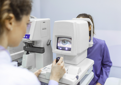
The main methods are corneal topography and tomography. They create a detailed map of the anterior surface of the eye and show areas of thinning and bulging. This allows for confirmation of the diagnosis and monitoring of progression.
In the early stages, such changes are only visible on instruments and are not noticeable during a routine examination. Therefore, without these tests, the disease can easily be missed.
Pachymetry, keratometry and refractive testing
Pachymetry is used to measure the thickness of the cornea. Keratometry measures its curvature. Refraction testing explains how the eye focuses light and why vision becomes unstable. The doctor compares these data and sees how much the cornea has changed.
A single examination is often not enough. It's important to compare results over time. This way, the doctor can determine whether the disease is stagnant or progressing, and can choose the next course of action without unnecessary prescriptions.
Keratoconus treatment and management methods
| Method | What it solves | When it's suitable | Advantages |
|---|---|---|---|
| Glasses and soft lenses | Primary correction | Early stages | Easy start |
| Rigid and scleral lenses | Improving vision quality | Irregular astigmatism | Precise correction |
| Crosslinking | Corneal stabilization | Progression | Helps slow the disease |
| Corneal rings | Shape correction | As indicated | Improving optics |
Glasses, soft and hard contact lenses
In the early stages, glasses and soft contact lenses are helpful. When astigmatism becomes irregular, hard contact lenses and scleral lenses work best. They create a smooth refractive surface and significantly improve clarity.
Fitting these lenses is more complicated than usual and requires the doctor's experience. However, the visual results are often noticeably better than with glasses.
Crosslinking as a method for corneal stabilization
Crosslinking is used to stop progression. This method strengthens the bonds within the cornea and makes it more stable. It does not restore the cornea's original shape, but it helps slow the disease at an early stage.
The earlier crosslinking is performed, the greater the chance of preserving current vision. Therefore, this method is especially valuable at the first signs of progression.
Corneal rings and surgical treatment according to indications
In some cases, corneal rings are implanted into the corneal layer to even out its shape. For severe changes, other surgical methods are considered, including corneal transplantation. Surgery for keratoconus is considered when conservative correction is no longer effective.
General information
Our advantages in keratoconus diagnosis and treatment
Accurate diagnosis and monitoring of progression are essential for this disease. We combine modern equipment with clear support. Below is what the patient receives.
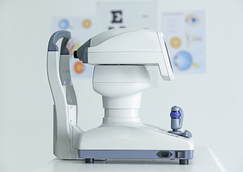
Modern equipment and accurate diagnosis
We assess the shape and thickness of the cornea using modern equipment. This examination helps detect keratoconus at an early stage and accurately monitor changes over time. The earlier the diagnosis, the greater the opportunity to choose a gentle approach and preserve vision.
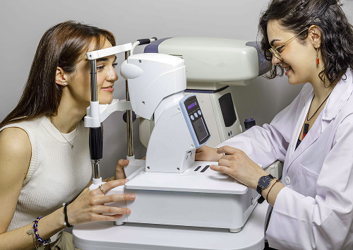
During the appointment, the doctor shows the patient the examination records and explains the changes visible in the diagnostic data. This makes it clearer why follow-up visits, corrections, or treatment are needed. We answer questions without rushing, so the patient understands every step and can make an informed decision.
Individualized treatment plan tailored to the stage of the disease
We select the treatment plan based on the stage and rate of progression. For some, correction is sufficient, while others require crosslinking or specially designed lenses. We make the decision together with the patient.
Clear explanations and dynamic monitoring
We explain the diagnosis in simple terms and show changes on corneal maps. We schedule follow-up visits to detect progression early. This monitoring helps ensure the right opportunity for stabilization is not missed.

FAQ
Below are short answers to questions that patients most often have before a consultation.
What is keratoconus?
Is it possible to stop keratoconus without surgery?
What tests are needed to confirm keratoconus?
When is keratoconus surgery necessary?
Category of Clinical Guidelines of the Ministry of Health of the Russian Federation — https://cr.minzdrav.gov.ru/
Ministry of Health of the Russian Federation — https://minzdrav.gov.ru/
EyePress — Portal of Russian Ophthalmology — https://eyepress.ru/
Russian Medical Journal — https://www.rmj.ru/
Media Sphere — https://www.mediasphera.ru/
Our doctors





















This award is given to clinics with the highest ratings according to user ratings, a large number of requests from this site, and in the absence of critical violations.

This award is given to clinics with the highest ratings according to user ratings. It means that the place is known, loved, and definitely worth visiting.

The ProDoctors portal collected 500 thousand reviews, compiled a rating of doctors based on them and awarded the best. We are proud that our doctors are among those awarded.
Make an appointment at a convenient time on the nearest date
Price
Other services

Appointment to the doctor
Reviews





















Our clinics


Application “Personal Account K+31”












Why keratoconus requires early diagnosis
Normal corneas are smooth and spherical, focusing light correctly. Keratoconus causes the cornea to thin, bulging into a cone. Light rays are refracted unevenly, blurring vision. This is initially subtle, but becomes more pronounced over time. Conventional vision corrections are unable to cope with such an uneven surface.
The danger is that, without monitoring, the disease can progress and significantly reduce vision. In the early stages, it is easier to keep it under control. In advanced cases, only complex treatment can help. Therefore, it is important not to ignore the early signs. Timely diagnosis is essential.