Orbitotomy in Moscow
Orbitotomy in Moscow is a surgical procedure. It is used to access the orbital tissues, which is necessary when the pathological process is located not on the surface of the eye, but deeper, in the orbital region. We consider this procedure only when indicated, after an in-person examination, imaging, and risk assessment, as the optic nerve, extraocular muscles, blood vessels, and other important structures are located nearby.
At K31, we guide patients from the initial consultation to recovery: we clarify the diagnosis, plan the scope of the procedure, choose the approach, and support them during the recovery period.

specialists

equipment

treatment


What is orbitotomy and when do we recommend it?
The orbit, or eye socket, is the area containing the eyeball, muscles, nerves, blood vessels, fatty tissue, and lacrimal gland. When a pathological lesion is located within this area, a simple superficial intervention is insufficient. In such cases, the doctor may consider orbitotomy as a way to carefully access the area.
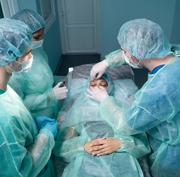
Orbitotomy as a Surgical Access to the Orbital Tissues
This specialty is related to ophthalmic surgery and requires experience working with the orbit. The intervention may involve oculoplastics, neuro-ophthalmology, maxillofacial surgery, or ENT referral if the process affects adjacent anatomical areas.
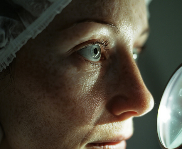
When Orbitotomy Helps Preserve Ocular Function and Quality of Life
The patient may experience double vision, pain, limited ocular movement, swelling, displacement of the eyeball, exophthalmos, or decreased vision. In such situations, surgery can be important not only from an aesthetic but also from a functional perspective.
Our goal is to eliminate or reduce the cause of the compression, obtain an accurate diagnosis, and preserve the maximum possible ocular function.
Main indications: neoplasms, biopsy, foreign bodies, decompression
The decision is made by the doctor after an examination, analysis of complaints, and CT or MRI data.
When we may recommend intervention:
- Suspected orbital neoplasm
- Need for orbital biopsy
- Removal of a foreign body
- Compression of orbital tissue
- Preparation for further treatment
- Orbital decompression in cases of severe exophthalmos or optic nerve compression
In some cases, removal of the neoplasm is required Orbitotomy is a common procedure, but sometimes the primary goal is to obtain material for morphological examination, relieve tissue pressure, or clarify the nature of the process.
How We Perform Orbitotomy in K31
Treatment is a step-by-step process, and here's how it works in our clinic.
Initial Consultation and Diagnosis
During the initial consultation, the ophthalmic surgeon clarifies the patient's complaints, duration of symptoms, previous surgeries, injuries, systemic diseases, and results of previous examinations. We assess vision, eyeball position, ocular motility, condition of the eyelids, conjunctiva, fundus, and signs of optic nerve involvement.
Sometimes the patient already has a diagnosis, but it is important for us to see the images themselves, as the size of the lesion, its depth, and its relationship to muscles, nerves, vessels, and bony walls are crucial for orbital surgery.
CT and MRI of the Orbits: Why Are They Necessary Before Surgery?
CT of the orbits helps assess bone structures, the consequences of trauma, the location of the foreign body, and the relationship of the lesion to the orbital walls. MRI of the orbits better depicts soft tissues, muscles, the optic nerve, vascular features, and the extent of the process.
Sometimes these methods complement each other. We use imaging to plan a safe approach, choose the surgical side, assess the extent, and discuss the prognosis.
How We Choose the Approach and Extent of Intervention
In some cases, an anterior approach is appropriate, while in others, a lateral approach, a medial approach, or an endoscopic approach through adjacent anatomical zones is required.
| Access type | When to choose | Pros | Features |
|---|---|---|---|
| Anterior | For superficial localization | Accuracy, accessibility | Depends on the intervention area |
| Lateral | For deep lesion location | Convenient visibility | Requires an experienced team |
| Endoscopic | When a minimally invasive approach is needed | Less tissue trauma | Not suitable in all cases |
In modern practice, orbital surgery strives for precision and reduced trauma, but a small incision is not always the best option.
Anesthesia and Surgical Stages
Anesthesia is selected individually. The choice is influenced by the extent of the procedure, the location of the lesion, the patient's age, any comorbidities, and the expected duration of the surgery.
During the procedure, the surgeon provides access to the orbital tissue, carefully isolates the desired area, and performs lesion removal, biopsy, foreign body removal, or decompression.
General information
Why patients choose K31 for orbitotomy in Moscow
A team of ophthalmic surgeons with experience in the orbit
The ophthalmic surgeon must have a thorough understanding of the anatomy of the orbit, possible lesion locations, and risks to vision. We carefully plan each procedure and discuss with the patient not only the procedure but also possible recovery scenarios.
The ophthalmic surgeon explains why the tumor needs to be removed, the alternatives available, the expected outcome, and any potential post-operative restrictions.
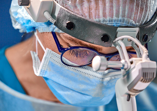
Modern equipment and gentle techniques
We use modern diagnostics and gentle surgical approaches when appropriate for the clinical situation.
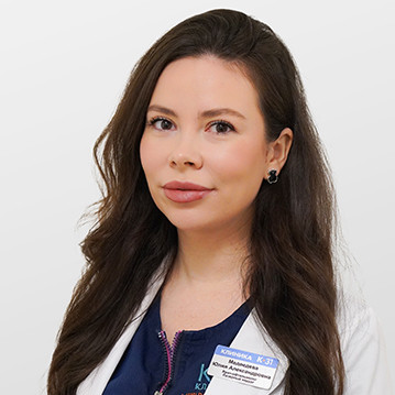
"In orbital surgery, it is especially important for us not just to remove the lesion, but to do so as precisely and gently as possible." "We evaluate the anatomy in advance, select the appropriate approach, and support the patient through every stage of recovery," says ophthalmic surgeon K31.
Individualized Treatment Plan and Safety Monitoring
Each orbital surgery requires a customized plan. We consider not only the imaging data but also the patient's complaints, symptom dynamics, vision, age, and comorbidities. If the procedure can be postponed for further examination, we do so.
We explain absolutely everything: what tests to take, what medications to approve, when to return, how the procedure will proceed, and what postoperative signs require contacting the doctor.
Postoperative Care and Observation
After the surgery, we schedule a follow-up examination, assessing healing, swelling severity, eye movement, diplopia, suture condition, and visual function. If necessary, we adjust recommendations and determine a timeframe for returning to normal activities.


Frequently Asked Questions
Is orbitotomy painful?
How long does recovery take?
Is inpatient care necessary after surgery?
When can I return to normal activities and work?
Sign up for a consultation at K31

How does the initial consultation proceed?
During the initial consultation, we clarify your complaints, conduct an examination, review your existing documentation, and explain whether further testing is necessary. If intervention is warranted, the doctor discusses possible access options, preparation, and expected recovery.
What tests should you bring?
It's helpful to bring CT and MRI results, previous ophthalmologist reports, surgical discharge summaries, laboratory results, and a list of your medications to the consultation.
Why it's best not to delay your consultation?
Pain, decreased vision, double vision, severe swelling, exophthalmos, or a suspected orbital mass require an in-person assessment. The sooner the doctor sees the patient and the imaging data, the sooner a safe approach can be chosen.
Schedule an appointment with K31 if you are recommended orbital surgery, need an orbitotomy in Moscow, or need a second opinion regarding an orbital mass. We will conduct a diagnosis, explain possible treatment options, and develop a plan that takes into account the patient's medical indications, safety, and quality of life.
Our doctors


This award is given to clinics with the highest ratings according to user ratings, a large number of requests from this site, and in the absence of critical violations.

This award is given to clinics with the highest ratings according to user ratings. It means that the place is known, loved, and definitely worth visiting.

The ProDoctors portal collected 500 thousand reviews, compiled a rating of doctors based on them and awarded the best. We are proud that our doctors are among those awarded.
Make an appointment at a convenient time on the nearest date
Price
Other services

Appointment to the doctor
Reviews


Our clinics

Application “Personal Account K+31”


