
Пневморетинопексия (пневматическая ретинопексия): лечение отслойки сетчатки
Проблемы со зрением часто развиваются незаметно, но существуют состояния, которые требуют немедленного медицинского вмешательства. Одно из таких опасных заболеваний — отслойка сетчатки, при которой нарушается питание светочувствительных клеток, что может привести к необратимой слепоте.
Современная офтальмология предлагает различные варианты помощи, среди которых выделяется пневморетинопексия — высокоэффективная и щадящая процедура. Этот малоинвазивный метод позволяет восстановить анатомическое положение тканей без проведения сложных полостных операций.
В клинике К+31 мы обеспечиваем полный цикл ведения пациента, начиная от экстренной диагностики и заканчивая тщательным контролем в реабилитационном периоде.

специалисты

оборудование

лечения






Что такое пневморетинопексия
В большинстве случаев проблема начинается с того, что возникает разрыв сетчатки, через который жидкость из стекловидного тела проникает под светочувствительную оболочку и отслоит ее от подлежащих сосудистых тканей. Пневморетинопексия — это амбулаторная процедура, направленная на закрытие этого дефекта изнутри с помощью микроинъекции специального газа.
Принцип метода: газовая тампонада и прижатие сетчатки
Офтальмохирург вводит внутрь глаза микроскопический объем специального вещества. Этот интраокулярный газ обладает свойством постепенно расширяться в первые дни после введения. Когда пациент принимает определенное положение тела, газовый пузырь всплывает и начинает оказывать направленное давление на зону повреждения.
Такая газовая тампонада выполняет сразу две важнейшие задачи:
- Она механически прижимает отслоившуюся внутреннюю оболочку обратно к сосудистой стенке глаза
- Газовый пузырь полностью блокирует разрыв сетчатки, не позволяя внутриглазной жидкости проникать под неё дальше
После того как оболочки сближаются, врач может надежно зафиксировать их, чтобы предотвратить повторное скопление жидкости. Постепенно газ в глазу самостоятельно рассасывается, а его место занимает естественная внутриглазная жидкость.
Чем пневморетинопексия отличается от других способов лечения
Традиционное лечение отслойки сетчатки часто ассоциируется с такими операциями, как витрэктомия или склеральное пломбирование. Эти методики требуют работы в условиях операционного блока, наложения швов и длительного наркоза.
Во время витрэктомии стекловидное тело удаляется практически полностью, а при склеральном пломбировании на глазное яблоко снаружи подшивают специальный силиконовый жгут. Пневматическая ретинопексия работает локально и прицельно, воздействуя непосредственно на причину отслоения.
Когда рекомендуют пневморетинопексию
Данный способ лечения имеет четко очерченные границы применения, поскольку он не является универсальным средством для всех видов глазных патологий.
Основные показания к процедуре
Процедура эффективна, если дефект расположен в верхней половине глазного дна — примерно от 8 до 4 часов по условному циферблату. Это связано с тем, что газовый пузырь всегда стремится вверх, и обеспечить адекватное давление на нижние отделы глаза технически невозможно.
В каких случаях метод особенно эффективен
Максимальную результативность процедура демонстрирует в следующих ситуациях:
- При свежем разрыве сетчатки, который произошел недавно и еще не вызвал обширных фиброзных изменений
- При небольшом объеме отслойки, когда пораженная зона локализована и не затрагивает центральную область видимости
- Если важно минимально травмировать ткани глаза, например, у пожилых пациентов или людей с тяжелыми сопутствующими соматическими заболеваниями
- Если пациент может соблюдать положение головы после процедуры, понимая важность этого этапа
- Если врач видит хорошие шансы на успех именно при таком подходе после проведения ультразвукового исследования глаза
Когда лучше рассмотреть альтернативы
Пневматическая ретинопексия окажется неэффективной, если разрыв сетчатки локализуется в нижних отделах глазного бокала, так как газ просто не сможет прижать эту зону. Также метод не используют при наличии множественных дефектов в разных квадрантах глаза. В таких запущенных случаях стандартом лечения становится полноценная витрэктомия, в ходе которой хирург убирает измененное стекловидное тело и расправляет ткани изнутри с помощью тяжелой воды или силиконового масла. Если же обнаружен изолированный дефект без скопления жидкости под ним, то может быть проведена только лазерная коагуляция сетчатки без введения газовой смеси.
Как проходит процедура
Знание того, как устроена процедура, помогает снизить уровень тревоги и подготовиться к лечению.
Подготовка к пневморетинопексии
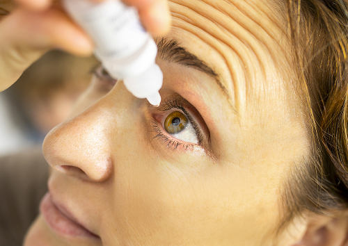
Перед процедурой пациент проходит стандартный осмотр, офтальмолог расширяет зрачок с помощью специальных капель и обрабатывает кожу вокруг глаза и конъюнктивальную полость антисептическим раствором для обеспечения стерильности.
Основные этапы проведения
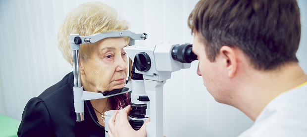
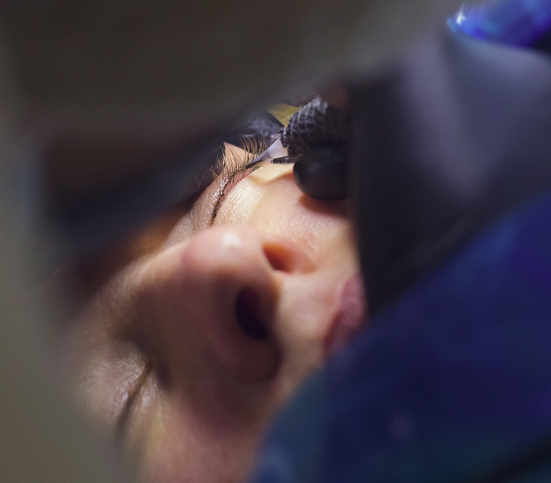
Пациент располагается в операционном кресле. Хирург использует специальный микроскоп для точной визуализации внутренних структур. Через тонкую иглу в полость глаза вводится подготовленный интраокулярный газ в строго рассчитанной дозировке. В момент введения человек может заметить появление темного облака или плавающего шара в поле зрения — это абсолютно нормально и свидетельствует о том, что газовый пузырь оказался в нужной зоне. Сразу после инъекции врач измеряет внутриглазное давление, чтобы убедиться в безопасности проведенной манипуляции.
Что происходит после введения газа
После того как игла извлечена, а давление стабилизировано, выполняется лазерная коагуляция сетчатки вокруг зоны разрыва. Иногда лазерное лечение переносят на несколько дней вперед, давая газу время полностью расправить и прижать ткани.
Лазерные лучи создают точечные микроожоги по краям дефекта. В процессе заживления этих точек образуются прочные спайки, которые намертво прикрепляют сетчатку к подлежащим тканям, исключая повторное проникновение жидкости.
Общая информация
Восстановление после пневморетинопексии
Период после процедуры требует от пациента высокой дисциплины, поскольку именно на этом этапе закладывается итоговый результат лечения.
Режим и положение головы после процедуры
Как только газ в глазу начинает расширяться, пациенту необходимо принять специфическое положение тела. Это называется позиционирование головы, и его характер зависит от того, где именно находился разрыв. Задача заключается в том, чтобы газовый пузырь постоянно всплывал точно в направлении повреждения и удерживал его в прижатом состоянии. Такого режима необходимо придерживаться в течение первых нескольких суток, включая время сна.
Ограничения в восстановительный период
Правильный послеоперационный режим включает полный отказ от тяжелых физических нагрузок, подъема тяжестей и резких наклонов туловища вниз.Не трите прооперированный глаз руками и избегайте попадания в него воды или мыла при умывании. Пока газовый пузырь остается в глазу, категорически запрещены авиаперелеты и поездки в горы. Перепады атмосферного давления вызовут резкое расширение газа, что может спровоцировать опасный скачок внутриглазного давления и необратимую потерю зрения.
Когда обычно появляется эффект и что отслеживать
Первые признаки улучшения пациент замечает уже в первые дни после процедуры, когда спадает отек и прижатая оболочка начинает снова воспринимать свет. По мере того как происходит восстановление зрения, серая пелена или занавес перед глазом начинают уменьшаться. Однако итоговое качество видимости будет понятно только после полного рассасывания газа.

Часто задаваемые вопросы
Больно ли делать пневморетинопексию?
Сколько длится восстановление после процедуры?
Можно ли летать на самолете после введения газа?
Что будет, если не соблюдать рекомендации врача?
Рубрикатор клинических рекомендаций Минздрава РФ — https://cr.minzdrav.gov.ru/
НМИЦ МНТК «Микрохирургия глаза» им. акад. С. Н. Фёдорова — https://www.mntk.ru/
НМИЦ глазных болезней им. Гельмгольца — https://www.helmholtzeyeinstitute.ru/
eLIBRARY.RU — https://elibrary.ru/
CyberLeninka — https://cyberleninka.ru/
Наши врачи




















Такая награда выдаётся клиникам с самым высоким рейтингом по оценкам пользователей, большим количеством обращений с данной площадки, при отсутствии критичных нарушений.

Такая награда выдается клиникам с самым высоким рейтингом по оценкам пользователей. Она означает, что место знают, любят и сюда точно нужно зайти.

Портал ПроДокторов собрал 500 тысяч отзывов, составил по ним рейтинг врачей и наградил лучших. Гордимся, что среди награжденных есть наши врачи.
Запишитесь на прием в удобное время на ближайшую дату
Стоимость
Другие услуги

Запись к врачу
Отзывы





















Наши клиники


Приложение «Личный кабинет К+31»












Преимущества пневморетинопексии для пациента
Выбор этого метода обусловлен его высокой эффективностью при минимальной нагрузке на организм.
Малоинвазивность и щадящее воздействие
Процедура не требует госпитализации в стационар и проводится под местной капельной анестезией, что исключает токсическое воздействие общего наркоза на сердце и сосуды. Пациент не чувствует боли, а само вмешательство занимает всего 15–20 минут.
Сохранение анатомии глаза и точечное воздействие
Во время манипуляции стекловидное тело не удаляется, сохраняются все естественные структуры и гидродинамика глаза. Врач воздействует исключительно на проблемную зону. Поскольку сетчатка глаза не подвергается грубому механическому давлению инструментами, снижается риск развития послеоперационного воспаления и отдаленных осложнений, таких как вторичная глаукома или ускоренное развитие катаракты.
Наши преимущества: точная диагностика, контроль и сопровождение
Мы используем оптическую когерентную томографию экспертного класса и ультразвуковое сканирование высокого разрешения, что позволяет выявить даже самые мелкие и скрытые разрывы. Наша команда, в которую входят опытные диагносты и витреоретинальные хирурги, сопровождает пациента на каждом этапе, от момента первого обращения с острыми симптомами до полного прирастания тканей.