Vitreoretinal surgery in Moscow
Vitreoretinal surgery is necessary for diseases of the retina and internal structures of the eye. The doctor works with delicate tissues that affect clear vision. Therefore, the decision to perform surgery is made only after diagnosis.
At "K+31," patients are treated by ophthalmologists who specialize in retinal diseases. During the examination, we evaluate the retina, vitreous body, and macula—the central area responsible for clear vision—as well as potential risks. If retinal surgery is indicated, the doctor thoroughly explains the diagnosis, treatment plan, and post-operative restrictions.

specialists

equipment

treatment


For what diseases and complications do we recommend surgery?
Indications are determined by an ophthalmic surgeon after examination and tests. The doctor looks at the central zone of the retina, its periphery, transparency of the media, the state of the vessels and the duration of the process. Old conclusions are useful, but the decision is made based on fresh diagnostic data.
Most often, surgical treatment is discussed in the following conditions:
- Retinal detachment or high risk of its development
- Macular hole, when the central zone of the retina is damaged
- Hemophthalmos, that is, bleeding into the eye
- Epiretinal membrane, which tightens the surface of the retina
- Diabetic retinopathy with complications
- Traction retinal detachment, when scar tissue pulls the retina
After the diagnosis, we explain to the patient why it is possible to observe or why it is better not to postpone the surgery.
Retinal detachment
With retinal detachment, the retina loses its normal attachment to the inner tissues of the eye. This is often preceded by flashes of light, a sudden increase in floaters, a shadow, a "curtain," or a veil in front of the eye. Vision may deteriorate gradually, but sometimes it changes rapidly. These symptoms require immediate consultation.
In this condition, retinal surgery aims to restore the tissue to its proper position. The prognosis depends on whether the macula is affected, how long it has been since the symptoms began, and whether there are any tears. The sooner the patient sees a doctor, the more accurate the treatment plan can be.
Macular hole
A macular hole is a defect in the central part of the retina. The macula is responsible for reading, facial recognition, and working with fine details. When damaged, lines may become distorted, and a spot appears in the center of the visual field.
With this diagnosis, vitrectomy helps relieve tension from the vitreous. Internal support is often used at the end of the surgery to better align the edges of the hole. A gas bubble is often used during macular hole surgery to support the edges of the defect during healing.
Hemophthalmos and vitreous hemorrhage
Hemophthalmos is a hemorrhage into the eye. The blood can partially or completely obscure vision, causing the patient to see dark spots, cloudiness, or a sharp decrease in vision. A doctor cannot always clearly examine the retina through a dense hemorrhage.
If the blood does not resolve over time, or the doctor suspects a tear, detachment, or vascular complication, surgery may be necessary. During the procedure, a retinal surgeon removes the blood and examines the retina from the inside. If there are any dangerous areas, they are treated with a laser.
Epiretinal membrane and traction
The epiretinal membrane forms on the surface of the retina and can pull on its central part. This causes distorted lines, difficulty distinguishing fine details, and fatigue when reading. The greater the tension, the more noticeable the change in vision.
If the changes are severe, the doctor may recommend retinal surgery. The surgeon removes the membrane and reduces the tension in the macular area. The result depends on how long the process has been going on, so rapid improvement is not always achieved. The doctor discusses these timeframes with the patient before the procedure.
Diabetic retinopathy and tractional retinal detachment
Diabetic retinopathy damages the retinal blood vessels. In severe cases, the following may occur:
- Hemorrhages
- Scar tissue
- Tension zones
These scar bands can pull the retina and change its position.
Traction retinal detachment develops precisely because of this tension. In this situation, vitrectomy is necessary to remove blood, adhesions, and pathological traction. Without treatment, the risk of permanent vision loss is higher.
General information
How we perform vitreoretinal surgery
Before the procedure, we conduct an examination and choose a safe route. It's important to understand not only the diagnosis but also the condition of the entire eye. This determines the extent of the surgery, the type of anesthesia, the method of internal support, and the postoperative regimen.
Diagnosis and preparation for the surgery
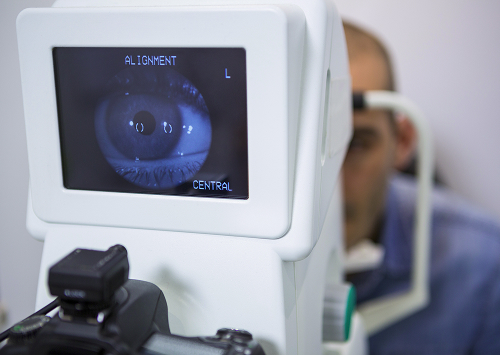
Before treatment, a retinal diagnosis is necessary. We assess visual acuity, the macula, the periphery, the transparency of the media, and areas of tension. We also consider chronic diseases, previous surgeries, and the medications the patient is taking.
The examination may include:
- Includes OCT
- Fundus examination
- Retinal photography
- Ultrasound of the eye
OCT shows the thin layers of the macula. Ultrasound is helpful if blood or dense clouding prevents the doctor from seeing the fundus.
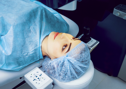
How is the surgery performed?
The procedure is performed in the operating room. The ophthalmic surgeon works through small incisions, inserts microinstruments, and removes the abnormal contents of the eye. The doctor then removes blood, membranes, traction, and other tissues that are pulling on the retina.
For tears or risk areas, endolaser coagulation may be used. It helps treat the retina from the inside. At the end of the surgery, the doctor chooses how to support the retina: with a solution, gas tamponade, or silicone oil.
The doctor chooses the method of internal support based on the diagnosis and the condition of the retina. Gas dissipates on its own over time, but while it is in the eye, the patient must strictly adhere to the regimen. Silicone oil lasts longer, so in some cases it is removed later in a separate procedure.
Recovery after vitrectomy
After surgery, the patient receives a prescription for eye drops, restrictions, and follow-up appointments. During the postoperative period, the following is prohibited:
- Changing treatment on your own
- Rubbing your eye
- Suddenly increasing your workload
- Changing your head position
If gas is present in the eye, flying is prohibited until cleared by the doctor.
Vision recovery is gradual. The outcome is influenced by the diagnosis, the condition of the macula, the duration of the injury, vascular changes, and adherence to the regimen.
We do not promise the same results for all patients, as the initial condition of each eye is different.
| Tamponade method | When to use | Recovery features |
|---|---|---|
| Gas tamponade | Retinal detachment, macular hole | Head position must be maintained flying is prohibited until the gas has completely resolved. |
| Silicone oil | Severe detachments, high risk of recurrent retinal displacement | Re-operation to remove the oil may be required. |
| Balanced salt solution | Hemophthalmos, intact epiretinal membrane | Usually does not require head positioning as with gas. |
After discharge, the patient remains under observation. Follow-up is necessary for the doctor to assess the retina, intraocular pressure, and tissue response to treatment.

FAQ
What is vitreoretinal surgery and how does it differ from vitrectomy?
When is surgery urgent?
How safe is vitreoretinal surgery?
You should schedule an appointment if you have already been diagnosed, have sudden symptoms, or have a referral from an ophthalmologist. Bring the following:
- Test results
- Discharge reports
- Fundus images
- Medication list
- Chronic disease information
This will help you quickly understand your medical history.
During the consultation, we will conduct an examination, prescribe tests, and explain the treatment plan. If surgery is not necessary, the doctor will advise on how to monitor your condition. If surgery is indicated, you will receive a preparation plan, timeframe, restrictions, and a clear postoperative care plan.
Our doctors


This award is given to clinics with the highest ratings according to user ratings, a large number of requests from this site, and in the absence of critical violations.

This award is given to clinics with the highest ratings according to user ratings. It means that the place is known, loved, and definitely worth visiting.

The ProDoctors portal collected 500 thousand reviews, compiled a rating of doctors based on them and awarded the best. We are proud that our doctors are among those awarded.
Make an appointment at a convenient time on the nearest date
Price
Other services

Appointment to the doctor
Reviews


Our clinics

Application “Personal Account K+31”


What is vitreoretinal surgery and when is it needed?
Vitreoretinal surgery is a subspecialty of ophthalmology that deals with surgeries on the retina and vitreous body.
The vitreous body is a transparent, gel-like structure inside the eye. Due to hemorrhage, inflammation, injury, or age-related changes, it can become cloudy, pulling on the retina, or preventing the doctor from accessing the damaged area.
Vitrectomy is one of the main methods in this field. During the procedure, the ophthalmic surgeon removes abnormal areas, blood, membranes, or strands that are damaging the retina. The goal of treatment is:
Surgery is not always necessary. Sometimes, the doctor chooses observation, treatment of inflammation, or laser treatment. But if there's a tear, severe hemorrhage, retinal tension, or a risk of retinal detachment, waiting is dangerous. In such situations, delay can worsen the prognosis.