
Iris melanoma (iris melanoma): symptoms, diagnosis, and treatment
When a new spot or nodule appears in such a prominent area of the eye, it always causes great anxiety. Our team deals with such patient concerns daily, so we strive to provide the clearest and most reliable information possible. Changes in pigmentation or structure of the anterior segment of the eye can indicate a serious pathology, among which iris melanoma occupies a special place. This rare but aggressive iris tumor requires immediate attention from qualified specialists.

specialists

equipment

treatment






Why does iris melanoma develop? Risk factors

Heredity and pigmentation characteristics
Modern medical science cannot yet identify a single, definitive cause that triggers this pathological process. However, long-term clinical observations have identified a group of factors that significantly increase the risk of developing this tumor. A person's natural pigmentation plays a significant role, as statistics show that people with fair skin and blue, gray, or green eyes are significantly more likely to experience this condition. Genetic predisposition and a strong family history are also considered by doctors.
UV radiation, trauma, and other possible triggers
Chronic exposure to sunlight without protective lenses can damage the DNA of iris cells and provoke their malignant transformation. Ophthalmic oncology also associates possible triggers with previous severe eye injuries or chronic inflammatory processes in the anterior segment. Sometimes the trigger is a simple pigmented lesion that existed for many years as a normal birthmark, but under the influence of external factors, transformed into a malignant nodule.
Symptoms that require a visit to the doctor
A spot, nodule, or darkening on the iris
The first obvious sign is often the appearance of a dark spot, nodule, or general darkening of the tissue. The patient notices that the usual eye pattern has become uneven, and the pigmented lesion itself gradually increases in size or changes its shape. Our team always emphasizes to patients that any new elements or rapid growth of existing spots require immediate professional evaluation.
Pupil deformation and iris color change
As iris melanoma grows, it begins to invade the surrounding muscular structures responsible for narrowing and dilating the pupillary opening. This process results in visible deformation of the pupil; it loses its round shape, becomes elongated, or shifts toward the affected side. Simultaneously, patients or their loved ones may notice a localized change in iris color, with one eye appearing darker than the other or acquiring an unusually muddy hue.
Pain, redness and decreased vision
Patients begin to complain of a gradual decrease in vision, along with the appearance of a fog or veil before their eyes, which is associated with a loss of optical transparency. When the tumor causes an accompanying inflammatory process, severe redness of the eye develops, often accompanied by discomfort. Eye pain itself usually indicates that the process has progressed far enough to affect the nerve endings or cause serious intraocular complications.
Increased intraocular pressure and discomfort
Tumor cells or the tumor itself can clog the eye's drainage system, which drains fluid. This leads to a sharp increase in intraocular pressure, causing a feeling of fullness, constant discomfort, headaches on the affected side, and the appearance of rainbow-colored rings when looking at light sources.
Below are warning signs to look out for:
- A new dark spot or a large nodule has appeared on the iris.
- An old pigmented spot has begun to rapidly increase in size.
- The pupil has lost its round shape or stopped reacting normally to light.
- The overall color of the iris in one eye has changed.
- Unexplained pain, redness, or decreased vision have developed.
How we diagnose iris melanoma
Ophthalmic oncologist examination and biomicroscopy
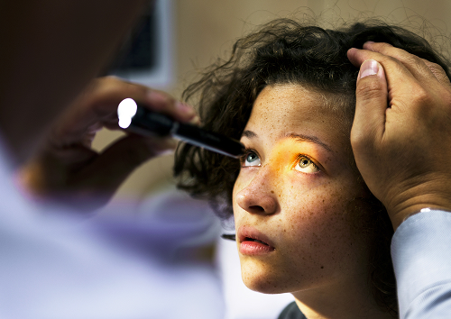
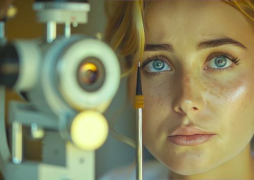
Since this pathology is malignant, a routine examination by a general ophthalmologist is often insufficient; a specialized consultation with an ophthalmic oncologist is necessary. At the K+31 clinic, the diagnostic process begins with a detailed interview and examination. The primary method for initial tissue assessment is biomicroscopy—an examination of the anterior segment of the eye using a slit lamp under high magnification.
Gonioscopy, eye ultrasound, OCT, and photofixation
To obtain a complete clinical picture, we always use high-tech instrumental examination methods:
- Gonioscopy — examination of the angle of the anterior chamber of the eye using a special lens, which is necessary to check whether the tumor has grown into the drainage system.
- Eye ultrasound — high-resolution B-mode ultrasound helps determine the exact size of the tumor, its thickness, and the extent of its spread into the deep layers.
- OCT — optical coherence tomography of the anterior segment provides layer-by-layer images of the iris microstructure with an accuracy of several microns.
- Photofixation — obtaining high-quality images is mandatory Digital iris imaging for precise monitoring of changes during subsequent visits.
How to distinguish melanoma from a nevus and other growths
The most important task in melanoma diagnosis is distinguishing malignant processes from benign changes. We need to conduct a thorough procedure, which in medicine is called differential diagnosis.
| Significance | Iris nevus | Iris melanoma |
|---|---|---|
| Growth of the lesion | Usually absent, stable condition | Characterized by obvious or gradual growth |
| Color and structure | Uniform color, clear borders | Uneven pigmentation, native vessels |
| Shape Pupil | Remains normal and round | Often deformed or displaced |
| Intraocular pressure | Always within normal limits | May increase if drainage is blocked |
| What to do | Monitoring | Specialized treatment as indicated |
General information
What treatment options are available?
Observation: when is it possible?
In some clinical situations, when dealing with a very small lesion and the physician is not absolutely certain of its malignancy, active surveillance may be chosen. The patient regularly visits a specialist, undergoes ultrasound examinations, and undergoes photographic documentation. If the size and structure of the nodule remain completely stable over a long period, active therapeutic interventions are not performed, as it may be a progressive iris nevus that does not require radical measures.
Organ-preserving treatment
Modern ophthalmic oncology has made enormous strides, so today we have virtually abandoned old, mutilating techniques. Organ-preserving treatment always takes precedence, the goal of which is the complete destruction of tumor cells while sparing as much healthy ocular tissue as possible.
Surgical Treatment and Follow-Up
If the tumor is too large to be treated with radiation, targeted surgical treatment is performed. This procedure, called an iridectomy, involves surgical excision of the affected area of the iris.
After any type of treatment, the patient must remain under strict monitoring, as there is a risk of metastasis to distant organs.

FAQ
How does iris melanoma differ from an iris nevus?
What symptoms should you be concerned about?
How is the diagnosis confirmed?
Is it possible to save the eye and vision?

Our doctors





















This award is given to clinics with the highest ratings according to user ratings, a large number of requests from this site, and in the absence of critical violations.

This award is given to clinics with the highest ratings according to user ratings. It means that the place is known, loved, and definitely worth visiting.

The ProDoctors portal collected 500 thousand reviews, compiled a rating of doctors based on them and awarded the best. We are proud that our doctors are among those awarded.
Make an appointment at a convenient time on the nearest date
Price
Other services

Appointment to the doctor
Reviews





















Our clinics


Application “Personal Account K+31”












What is iris melanoma?
This is a neoplasm that develops from pigment cells called melanocytes. The iris itself is rich in these cells, as they determine the individual color of our eyes, ranging from light blue to dark brown. When these cells malfunction, they begin to divide uncontrollably, resulting in the formation of ocular melanoma.
Malignant eye tumors can gradually destroy surrounding tissue, disrupt the circulation of intraocular fluid, and cause severe complications, so early, comprehensive diagnosis is crucial.