Eye biomicrography in Moscow: what the test shows and when it's needed
Eye biomicrography in Moscow not only helps with an examination but also captures accurate tissue images for subsequent comparison. This examination is especially important for inflammation, the consequences of trauma, chronic diseases, and during treatment monitoring. This examination provides an objective picture, stores the images in the clinic's archive, and uses them for dynamic monitoring.

specialists

equipment

treatment


What is biomicrophotography
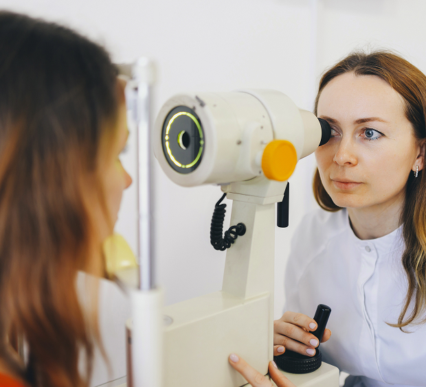
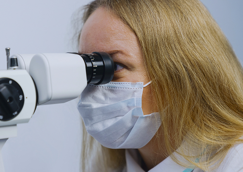
Biomicrography of the eye is a method of photographing structures using special optics and a digital camera connected to diagnostic equipment. Most often, examination is performed through a slit lamp. The doctor obtains an enlarged image of the tissue and stores the images in the medical records. This approach not only helps conduct an ophthalmological examination but also objectively assesses changes during follow-up visits.
Today, biomicrography of the eye in Moscow is used as part of modern vision diagnostics. This method eliminates the need to rely solely on a description of symptoms or a visual assessment during a visit. The images are stored in the clinic's archive and allow for comparison after several weeks or months.
During the procedure, the condition of the anterior segment of the eye, the cornea, iris, conjunctiva, and lens are analyzed. If necessary, images are taken at different magnifications and under different lighting angles.
How does biomicrography differ from a regular examination?
A routine examination only provides information about the patient's condition at the time of the visit. Biomicrography allows for additional image storage and use for further comparison. This is especially important in chronic conditions, after surgery, inflammation, and the consequences of eye trauma.
During a routine examination, the ophthalmologist verbally records changes in the medical record. A photograph of the eye helps preserve objective visual information. The doctor can review the images at any time and assess how the tissue condition is changing.
| Method | What it provides | When it is useful |
|---|---|---|
| Routine examination | Visual assessment of the condition | At the initial appointment |
| Biomicrography | Photographic recording and comparison over time | For treatment monitoring and documentation of changes |
What structures can be assessed in the image?
During an eye examination, a detailed image of the anterior segment tissues can be obtained. The images clearly show the cornea, iris, conjunctival vascular pattern, eyelid surface, and lens.
With digital photography, an ophthalmologist can identify signs of inflammation, microdamage, changes in tissue transparency, and the consequences of trauma. If a patient is undergoing treatment monitoring, the doctor compares current images with previous ones and evaluates progress.
Photographic documentation of the eye is used not only for diagnostic purposes but also for storing medical information.
When we recommend biomicrography
The procedure is prescribed in situations where it is important to document tissue condition and save examination results for future comparison. The examination is often included in a comprehensive ophthalmological examination and helps obtain a more accurate picture.
For inflammation, injury, and complaints of discomfort
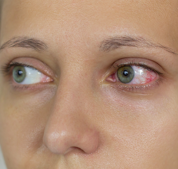
The doctor may recommend the procedure if you experience redness, a foreign body sensation, irritation, pain, photophobia, or deteriorating vision.
Biomicrography of the eye in Moscow is especially useful if you suspect eye inflammation, damage to the corneal surface, or the consequences of an injury.
When is biomicrography especially useful:
- For complaints of redness and irritation
- After an injury
- For treatment monitoring
- For monitoring chronic diseases
- Before a follow-up appointment to compare progress
For treatment and post-procedure monitoring
After prescribing therapy, it is important for the ophthalmologist to understand how the tissues are responding to treatment. In such situations, monitoring treatment based solely on the patient's complaints is insufficient.
A follow-up eye examination helps compare tissue condition before and after treatment. This is important after laser procedures, surgeries, therapy selection for inflammatory diseases, and during the recovery period.
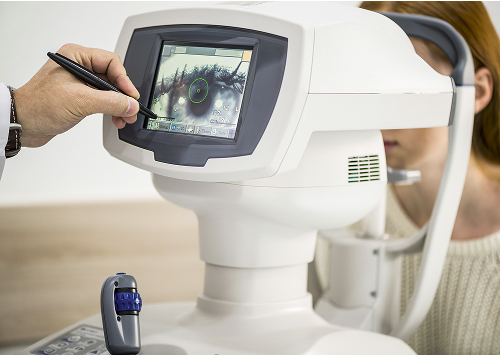
At the K+31 clinic, images are stored in an electronic system. This allows the ophthalmologist to quickly assess changes and adjust recommendations if necessary.
For follow-up monitoring
Some ophthalmological conditions require regular monitoring. In such cases, follow-up monitoring becomes an important part of treatment.
If a patient is undergoing long-term therapy or is seeing a doctor repeatedly, biomicrography helps objectively compare examination results. This reduces the likelihood of missing small changes that are difficult to notice during a routine examination.
General information
Advantages of biomicrography at K+31
At K+31, the examination is performed using modern equipment that allows for the production of detailed, high-quality images.
Photographic recording and storage of results
The main advantage of this method is the precise photographic recording of tissue condition. All images are stored in the clinic's archive and can be used for follow-up visits.
This photographic documentation of the eye helps assess changes objectively and not rely solely on the patient's description of symptoms.
Comparison of conditions over time
During follow-up visits, the ophthalmologist can open previous images and compare them with current results.
Dynamic monitoring helps detect changes early and evaluate the effectiveness of therapy. This approach makes treatment monitoring more accurate and understandable for the patient.
Comfortable reception and modern equipment
K+31 uses modern diagnostic systems and a digital slit lamp with image storage capabilities.
If necessary, you can quickly schedule a follow-up appointment with an ophthalmologist.
FAQ
What is biomicrography in simple terms?
Will it hurt?
Do I need to do any special preparation for the procedure?
When might a doctor prescribe this test?

Our doctors


This award is given to clinics with the highest ratings according to user ratings, a large number of requests from this site, and in the absence of critical violations.

This award is given to clinics with the highest ratings according to user ratings. It means that the place is known, loved, and definitely worth visiting.

The ProDoctors portal collected 500 thousand reviews, compiled a rating of doctors based on them and awarded the best. We are proud that our doctors are among those awarded.
Make an appointment at a convenient time on the nearest date
Price
Other services

Appointment to the doctor
Reviews


Our clinics

Application “Personal Account K+31”


How the study is conducted
In some cases, the ophthalmologist uses drops to dilate the pupil. After this procedure, temporary blurred vision and increased sensitivity to light are possible. For this reason, the patient may be uncomfortable driving immediately after the examination.
Procedure steps
The ophthalmologist uses equipment including a slit lamp and a digital camera. The patient is positioned in front of the device and maintains focus as instructed by the doctor.
The ophthalmologist sequentially evaluates the anterior segment of the eye, the condition of the cornea, iris, and lens. A series of images are then taken at varying magnifications. All images are saved in the medical records.
How long does biomicrography take and is there any discomfort?
The procedure typically takes from a few minutes to half an hour. The duration depends on the scope of the examination and the need for additional images.
For most patients, eye biomicrography is painless. It's a non-invasive diagnostic method and doesn't involve contact with internal structures.