Vitreosinopharyngeal surgery in Moscow: when is surgery necessary and how we help restore vision
Vitreosinopharyngeal surgery in Moscow is performed for complex retinal changes, when dense scar tissue forms inside the eye. These scar tissue pulls on the retina and can lead to damage. Eye drops do not address the underlying cause, so we assess the risk and decide whether retinal surgery is necessary.
At "K+31," patients are cared for by a team of ophthalmologists specializing in retinal diseases. Our goal is to preserve the anatomy of the eye and support vision restoration as much as possible.

specialists

equipment

treatment


When is vitreosinophorectomy really necessary?
Vitreosinopharyngeal surgery isn't necessary for every complaint of floaters or decreased vision. We consider it when there's a mechanical cause within the eye that can't be addressed with medication.
Traction retinal detachment
Traction retinal detachment occurs when scar tissue begins to pull on the retina and change its position. A person may notice uneven lines, a spot in front of the eye, blurred vision, or a sensation of a "curtain."
In this situation, retinal surgery helps relieve the tension and protect the areas that affect vision. The doctor evaluates the prognosis based on the duration of the process, the condition of the macula, blood vessels, and optic nerve.
Proliferative vitreoretinopathy
Proliferative vitreoretinopathy is a condition in which dense membranes form on the retina and inside the eye. These membranes contract and again pull on the tissue. This can lead to recurrent retinal detachment or worsening of the condition after previous treatment.
In such cases, vitreoretinal surgery requires careful manipulation of the membranes and risk areas. The surgeon may use endolaser, tamponade, and other internal support methods.
Vitreous hemorrhage and other complications
A vitreous hemorrhage can obscure the retinal view and dramatically reduce vision. Sometimes the blood resolves, but if the hemorrhage is dense, the cause may persist.
Other indications include adhesions after trauma, complications of inflammation, diabetic changes, and dense opacities.
How we conduct diagnostics before surgery

A retinal diagnosis is mandatory before treatment. We evaluate visual acuity, the macula, peripheral areas, internal media transparency, and areas of tension.
Previous reports help us understand the progress, but we make a decision only after a fresh examination.
Vitreoretinal surgery examination
The vitreoretinal surgeon examines the eye with the pupil dilated, unless contraindicated. They evaluate the retina, vitreous body, membranes, blood, tears, and areas of detachment. At this stage, the patient receives an initial explanation of the treatment plan.
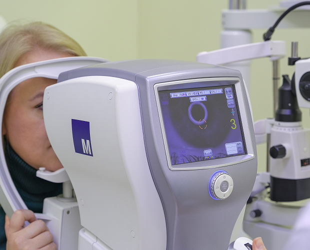
OCT, ultrasound, ophthalmoscopy, and other examinations
We select examinations based on the condition of the eye. Most often, we use OCT, ultrasound, ophthalmoscopy, fundus photography, and other methods as indicated. OCT helps evaluate the macula and the fine structures of the retina, and ultrasound is helpful when blood or opacities obscure the view.
This retinal diagnostic shows the doctor the areas of traction, the presence of detachment, and the severity of adhesions. Based on the examination results, the patient receives a clear treatment plan.
How we determine treatment strategy
We choose the treatment strategy after a complete eye assessment. The diagnosis, duration of changes, condition of the macula, transparency of the internal media, and concomitant diseases are important. We also consider previous surgeries and examine the other eye.
If the procedure is truly necessary, the ophthalmic surgeon will explain the steps, anesthesia options, and postoperative care in advance.
General information
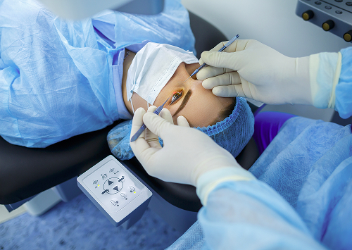
How is a vitreous wartectomy performed?
Vitreous wartectomy is performed in the operating room using microsurgical techniques. Through small incisions, the surgeon inserts fine instruments, removes damaged tissue, and reduces retinal tension. The procedure is performed near sensitive structures of the eye, so precision and careful technique are especially important.
Preparing for the procedure
Before the surgery, the patient undergoes an examination according to the clinic's plan. We review medications, allergies, chronic diseases, test results, and previous eye surgeries. We also explain what to do on the day of the procedure.
Main stages of the surgery
First, anesthesia is administered. Then, the surgeon makes microincisions and begins removing the vitreous humor to gain access to the retina. After this, the doctor separates the adhesions, removes the membranes, relieves tension, and removes any blood or opacities.
If necessary, an endolaser is used. It helps treat tears and risk areas from within the eye. Finally, the doctor selects a method of internal support.
What is tamponade and why is it needed?
Tamponade helps support the retina after surgery, ensuring it remains in the correct position. For this, the doctor chooses a gas mixture, gas tamponade, or silicone oil. The decision depends on the diagnosis, the location of the tears, and the risk of recurrent detachment. The gas gradually dissipates on its own, but during this period, it is important for the patient to adhere to the regimen. Silicone oil remains in the eye longer, so it is sometimes removed later as a separate step.


FAQ about vitreous wartectomy
Is the surgery painful?
How long does recovery take?
Is it possible to preserve vision after surgery?
What are the risks and limitations?
Vitreosinopharyngeal surgery in Moscow is performed after an in-person consultation and examination. We will evaluate the retina, vitreous body, macula, degree of traction, and any waiting periods. The doctor will then explain the next steps.
You can schedule a consultation in Moscow at "K+31" if you already have a diagnosis, referral, OCT results, or sudden symptoms. The sooner the doctor can see the retina, the more accurate the treatment plan.
When is it best not to delay your appointment?
Don't delay your appointment if you experience a sudden decrease in vision, flashes of light, a dark area in front of the eye, a sensation of a "curtain," a sudden increase in floaters, severe pain, or severe redness. These symptoms may be associated with conditions in which delaying treatment increases the risk of irreversible retinal damage.
What to bring to your appointment
Bring your passport, medical records, OCT and ultrasound results, fundus photographs, a list of medications, and information about your chronic illnesses. If you had retinal treatment, bring your previous reports.
Schedule a consultation in Moscow. We will conduct a diagnosis, explain your eye condition in simple terms, and develop a treatment plan that suits your diagnosis.
Clinical Guidelines of the Ministry of Health of the Russian Federation — https://cr.minzdrav.gov.ru/
Ophthalmology Bulletin — https://www.mediasphera.ru/journal/vestnik-oftalmologii
Russian Medical Journal — https://www.rmj.ru/
CyberLeninka — https://cyberleninka.ru/
EyePress - https://eyepress.ru/
Our doctors


This award is given to clinics with the highest ratings according to user ratings, a large number of requests from this site, and in the absence of critical violations.

This award is given to clinics with the highest ratings according to user ratings. It means that the place is known, loved, and definitely worth visiting.

The ProDoctors portal collected 500 thousand reviews, compiled a rating of doctors based on them and awarded the best. We are proud that our doctors are among those awarded.
Make an appointment at a convenient time on the nearest date
Price
Other services

Appointment to the doctor
Reviews


Our clinics

Application “Personal Account K+31”


What is vitreosinopharyngeal stenting?
Vitreosin-sinusectomy is a microsurgical procedure in which the surgeon removes:
These tissues can pull on the retina, deform it, and increase the risk of detachment. The procedure is part of vitreoretinal surgery and is performed under precise visualization.
Abnormal adhesions can appear after inflammation, hemorrhage, trauma, or diabetic changes. The greater the tension, the higher the risk of damage.
How does vitreosin-sinusectomy differ from vitrectomy?
Vitrectomy is the removal of abnormal vitreous humor. With adhesion changes, cleaning the environment alone is not enough, as the retina can be pulled by dense membranes. Therefore, the surgeon additionally works with the retinal bands.
Removing the vitreous provides access to the posterior segment of the eye. After this, the doctor can remove blood, separate the retinal bands, treat the risk areas with a laser, and perform internal retinal support.
For what conditions do we recommend this surgery?
The decision to perform the surgery is made by a vitreoretinal surgeon. After examination, they determine whether the central part of the retina is affected, the severity of the retinal band, how long the disease has been progressing, and the condition of the other eye.
Surgery is most often considered for the following indications:
After diagnosis, we explain to the patient why surgery or observation was chosen.