Peripheral Fundus Examination with a Goldmann Lens in Moscow
A peripheral fundus examination with a Goldmann lens is necessary when the doctor needs to see the peripheral areas of the fundus in detail and assess areas of the retina that may be difficult to see during a standard examination.
We view this examination not as a formal part of the appointment, but as an important step in vision diagnostics. This is especially relevant for patients with high myopia, complaints of flashing lights, floaters, eye injuries, and suspected changes that may not cause obvious symptoms for a long time.

specialists

equipment

treatment


When we recommend coming for an appointment
High Myopia and the Risk of Retinal Changes
With high myopia, the eye often has an altered shape, and the retina may be more vulnerable to stretching and peripheral dystrophies. Not every patient with myopia will experience dangerous changes, but we recommend that patients with high myopia have regular fundus examinations, even if their vision is stable and there are no complaints.
Floaters, Flashes, and a "Curtain" Before the Eyes
Floaters before the eyes may be associated with changes in the vitreous humor, but if they appear suddenly, it is important to rule out pathology. Flashes of light also require attention because they sometimes occur due to traction, when the vitreous humor exerts pressure on the retina.
If you experience a curtain, a shadow on the side, a sudden increase in opacities, or a deterioration in vision, we recommend an appointment as soon as possible.
Eye Trauma, Diabetes, Preparing for Surgeries
After an eye injury, it is important to ensure that the retina is intact, even if the eye appears normal. In diabetes, the doctor evaluates not only the central zones but also the periphery, as vascular changes can affect different areas of the fundus.
Before ophthalmological procedures, especially if lens surgery or laser vision correction is planned, the doctor may recommend a more extensive evaluation.
We especially recommend Goldmann lens examination for:
- Patients with high myopia
- Those who notice flashes of light or "floaters"
- After an eye injury
- If a tear or dystrophy is suspected
- Before ophthalmological procedures
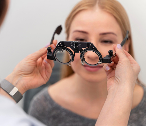
How is a Goldmann lens examination performed?
Preparing for the Procedure
No special preparation is usually required, but your doctor may use drops to dilate your pupils during your appointment. This dilation is called mydriasis. It allows for a better examination of the back of your eye and increases the effectiveness of the procedure.
If you wear contact lenses, you will need to remove them before the examination. It is also important to inform your doctor in advance about any glaucoma, allergies to eye drops, pregnancy, or eye injuries.
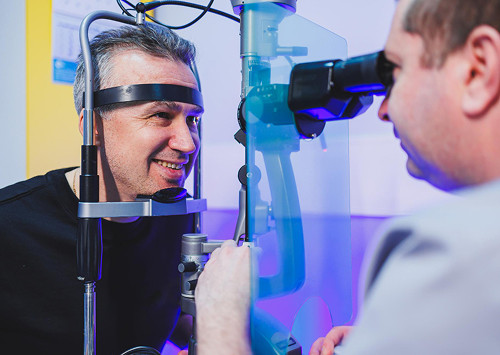
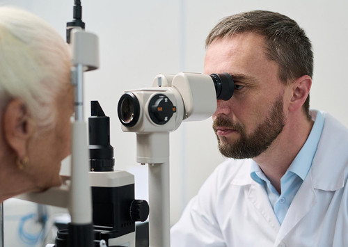
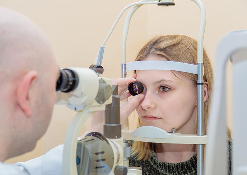
How the doctor conducts the examination
During the procedure, the patient sits at a slit lamp. The doctor administers anesthetic drops and then carefully places a diagnostic contact lens on the surface of the eye through a special medium. The doctor then directs the light and examines different areas sequentially.
The Goldmann lens requires precise technique, a calm pace, and sterility.
How long does the appointment last and what does the patient feel?
The examination itself usually takes a few minutes, but the full appointment takes longer because the doctor needs to collect the patient's complaints, perform a basic vision assessment, wait for the pupil to dilate if necessary, and then explain the results.
Mild discomfort from contact with the lens and bright light is possible.
General information
What changes can the examination reveal?
Peripheral retinal dystrophy
Peripheral retinal dystrophy can occur without obvious symptoms. Patients sometimes discover it accidentally, during a pre-operative examination or routine checkup.
During the procedure, the doctor evaluates the shape, location, and severity of the degenerative areas. Not every finding requires treatment.
Retinal Tears and the Risk of Detachment
A retinal tear can become a dangerous condition if fluid begins to leak under the retina. In this situation, the risk of retinal detachment increases, which requires urgent specialized care.
A second retinal tear may be located far in the periphery and not cause a noticeable decrease in central vision. Therefore, a detailed retinal diagnosis is especially important in cases of sudden flashes, numerous floaters, and trauma.
Associated changes in the vitreous
The vitreous body can change structure with age or myopia. Sometimes these changes are harmless, but in other cases they create tension in the attachment zone.

Frequently Asked Questions
Is a Goldmann lens examination painful?
Do I need to make any special preparations for the procedure?
Can I return to my normal activities immediately?
What should I do if I experience discomfort?
Why it is important not to delay diagnosis

Early Detection of Retinal Changes
Many changes do not interfere with vision for a long time. A person may be able to read, work, and drive well, but still have areas of thinning or degeneration that require monitoring. This is why a preventive examination is important not only for complaints.
A second preventive examination is especially necessary for patients at risk. These include those with high myopia, eye injuries, diabetes, a hereditary predisposition, and planned ophthalmological procedures.
How We Help Preserve Vision
We don't just conduct an ophthalmological examination; we also assess vision risks. If the retina is stable, the patient receives recommendations for monitoring. If there are suspicious areas, the doctor explains the next steps.
Our goal is to prevent complications and provide accurate diagnosis without unnecessary anxiety.
Book an eye examination in Moscow

When it's especially important to seek immediate attention
You should schedule a fundus examination in Moscow without delay if you experience new flashes, a sharp increase in floaters, a cloudy vision, decreased vision, or have suffered an injury.
We also recommend an examination before ophthalmological procedures, for high myopia, and for previously identified changes. If you're looking for an ophthalmologist in Moscow who can carefully evaluate your condition and explain the results, schedule a consultation at our clinic.
How to schedule an appointment
Submit a request on our website or choose a convenient time by phone. We'll advise you on any preparation needed, how much time to allocate for your visit, and whether you can plan any activities after your appointment.
A fundus examination helps you quickly detect changes that aren't always noticeable during a routine examination. Schedule an appointment if you need a retinal diagnosis, a detailed doctor's report, and a clear plan for next steps.
Our doctors


This award is given to clinics with the highest ratings according to user ratings, a large number of requests from this site, and in the absence of critical violations.

This award is given to clinics with the highest ratings according to user ratings. It means that the place is known, loved, and definitely worth visiting.

The ProDoctors portal collected 500 thousand reviews, compiled a rating of doctors based on them and awarded the best. We are proud that our doctors are among those awarded.
Make an appointment at a convenient time on the nearest date
Price
Other services

Appointment to the doctor
Reviews


Our clinics

Application “Personal Account K+31”


What is a fundus examination and why is it necessary?
This is an assessment of the peripheral areas where thinning, degenerative zones, and tears may form. These areas do not always cause pain or noticeable visual impairment, so the patient may not be aware of the risk until more worrisome symptoms appear.
This procedure is important for the physician because the periphery often requires contact ophthalmoscopy.
Why standard diagnostics are often insufficient
A standard fundus examination is effective in assessing the central areas, the optic disc, blood vessels, and some of the periphery, but the peripheral areas may remain difficult to access.
The patient may have fairly good vision, but areas of thinning or localized degeneration may already be present in the periphery. This is why, for certain complaints or risk factors, we expand the examination and conduct a more detailed diagnosis.
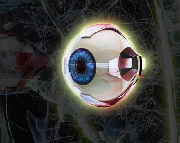
What can a Goldmann lens see?
A Goldmann lens is a contact diagnostic instrument used by a doctor with a slit lamp. This three-mirror lens allows for viewing different areas of the eye, including the peripheral areas.
A Goldmann lens allows for evaluation of the retina, identifying areas of thinning, suspicious areas, localized tears, and signs of vitreous tension.