
Blepharophimosis (Blepharophimosis Syndrome): Diagnosis and Treatment
Blepharophimosis changes the shape of the palpebral fissure and the position of the eyelids. Externally, this may appear as a cosmetic feature, but sometimes there is a functional defect: the upper eyelid obscures vision, and the child raises the chin.
We assess the condition in person: a photograph cannot determine muscle function and the impact on vision.

specialists

equipment

treatment






What is blepharophimosis?
Blepharophimosis syndrome is a condition in which the palpebral fissure is shortened horizontally and appears normal. This is often accompanied by:
- Dropping of the upper eyelids
- Fold at the inner corner
- Increased distance between the corners
For the doctor, the entire anatomy of the eyelids is important.
How is blepharophimosis different from regular ptosis?
Upper eyelid ptosis is a drooping eyelid that can be a separate problem. The doctor examines the extent to which the eyelid margin covers the pupil and obstructs vision. With blepharophimosis, other anatomical features are also present: a shortened palpebral fissure, an altered inner angle, and a lack of symmetry in the eyes.
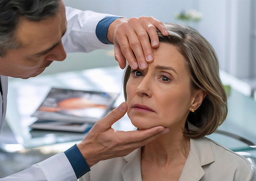
Therefore, during an appointment, not only eyelid height is assessed: the function of the levator muscle, the position of the eyebrows, and the impact of these changes on visual function are also examined.
What structures of the eyelids and the angle of the eye are affected?
With this syndrome, the upper and lower eyelids, the skin at the inner angle, and the medial ligament change. This changes the shape of the palpebral fissure. Epicanthus appears as a fold of skin at the bridge of the nose that partially covers the inner angle of the eye. Telecanthus means that the distance between the inner angles is increased, although the position of the eyeballs remains normal.
To help patients distinguish between similar conditions, we evaluate them based on several characteristics:
| Characteristic | What it looks like | How it affects vision | What the doctor does |
|---|---|---|---|
| Blepharophimosis | Narrow palpebral fissure, bilateral ptosis, fold | May narrow vision and cause head tilt | Planned comprehensive correction |
| Upper eyelid ptosis | Drooping eyelid, pupil covered | If the pupil is covered, there is a risk of amblyopia | Selects the lifting method |
| Epicanthus | Skin fold at the inner corner | Usually affects appearance | Considers plastic surgery according to indications |
The table does not replace an examination. It shows why the approach may vary.
Why does blepharophimosis occur?
Congenital blepharophimosis is associated with the developmental characteristics of the eyelids. In some cases, it is a hereditary syndrome. If there are family history signs, the doctor will confirm the patient's medical history and recommend further consultations if necessary.
Acquired Changes and Associated Causes
Acquired changes are less common, but it is important not to miss them. They may resemble blepharophimosis in appearance: the palpebral fissure narrows, the eyelid droops, and the inner corner appears different. However, the underlying cause may be different. Sometimes, the eyelid shape is altered by trauma, post-surgical scarring, prolonged inflammation, age-related tissue weakening, or nerve and muscle dysfunction.
During an appointment, the doctor will look beyond the patient's appearance. It is important to understand what exactly has changed and how it affects vision. Typically, the following are assessed:
- Upper eyelid position
- Width of the palpebral fissure
- Skin and scar condition
- Levator muscle function
- Eyelid closure
- Ocular surface
- Visual function
An examination is necessary to avoid confusing a congenital eyelid structure with an acquired problem. They may appear similar. But the causes are different, and the approach will also be different.
Therefore, the doctor doesn't rely solely on photographs. They take measurements, check eyelid movement, examine the cornea, and then decide whether correction, observation, or further examination is necessary.
When blepharophimosis requires special attention
If the palpebral fissure limits vision, delaying the examination is undesirable. The child may tilt their head back, close one eye, and have difficulty maintaining their gaze. In this case, a consultation is needed as soon as possible. In adults, pain, severe redness, and a sudden deterioration in vision should be a cause for concern.
General information
When to see a doctor
It's important to have your eyelids evaluated as early as possible in childhood. The eye must receive adequate visual stimulation, otherwise, vision development problems may develop.
A raised chin, squinting, and a tendency to turn or tilt the head when looking forward should be cause for concern. In this situation, an in-person examination is necessary, without relying on a photograph.
An ophthalmologist appointment is recommended if one or more of the following signs are present:
- The palpebral fissure is noticeably narrower than normal
- The upper eyelid partially covers the pupil
- The child tilts their head back to see better
- One eye appears smaller or narrower than the other
- There are complaints of visual discomfort
After the examination, the doctor explains the further treatment plan.
Complaints in Adults
In adults, the reasons for seeking an ophthalmologist include heavy eyelids, asymmetry, limited superior vision, and fatigue when reading and working at a screen. Sometimes a patient presents with an aesthetic complaint, but a functional defect is discovered during the examination. If necessary, the patient is examined by an ophthalmologist.
When a consultation should not be delayed
An ophthalmologist consultation is necessary without waiting if the eyelid covers the pupil or the child constantly raises the chin when looking forward. You should also consult a doctor immediately:
- In case of pain
- Noticeable redness
- Discharge
- A sharp decrease in vision
Self-prescribed eye drops and ointments can mask the symptoms, but they do not address the underlying anatomical disorder.
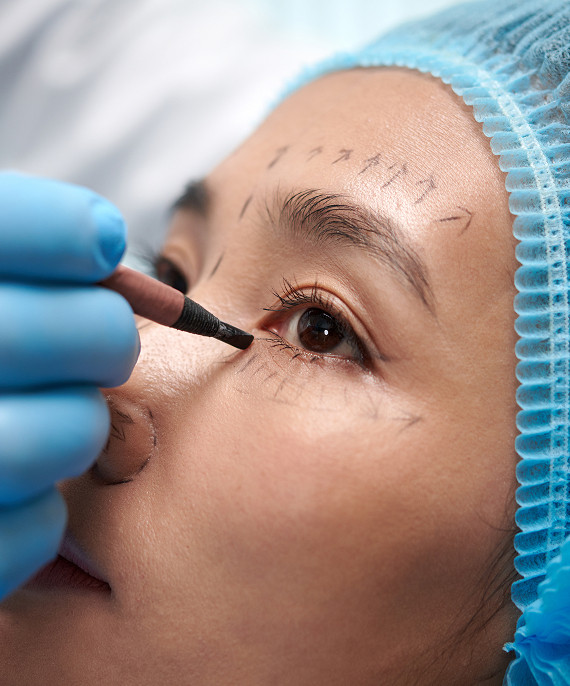
Blepharophimosis correction is indicated when narrowing of the slit, fold, and ptosis interfere with vision, cause a forced head position, or significantly disrupt symmetry. In children, the decision is made especially carefully due to the risk of affecting the development of the visual system. For minor changes, the doctor may opt for observation.
How the surgery is performed
The surgery may include several stages: widening the palpebral fissure, correction of the inner corner, and lifting the upper eyelid. If the epicanthal fold is pronounced, the doctor plans eyelid surgery. If the muscle is weak, a method for fixing or lifting the eyelid is selected. Sometimes, eyelid surgery is performed in stages to more accurately achieve shape and symmetry.
What results can be expected
Treatment is aimed at several goals at once: lifting the eyelids, expanding the field of view, and improving the shape of the ocular area. After blepharophimosis correction, a person often finds it easier to look forward without excessive forehead tension and the usual head tilt. The final result depends on the structure of the eyelids, the severity of ptosis, the patient's age, and individual tissue healing.
Frequently Asked Questions
What is blepharophimosis?
Can blepharophimosis syndrome be corrected without surgery?
What is the best age to seek a consultation?
What are the results of correction?
Conclusion

Blepharophimosis syndrome requires an in-person diagnosis. Sometimes the appearance of the eyelids is related to limited vision and strain on the visual system.
Therefore, we examine not only the shape of the eyelids but also the muscle function, the position of the inner angle, and the impact on everyday vision. This examination helps choose the right approach—observation or correction.
Congenital blepharophimosis in children is best assessed as early as possible. In adults, an ophthalmologist consultation is necessary if visual discomfort, asymmetry, and fatigue are present. An in-person consultation provides the patient with a clear diagnosis, a safe plan, and a calm and supportive approach.
References
- Clinical Guidelines Portal of the Ministry of Health of the Russian Federation
- CyberLeninka — Search by topic "блефарофимоз"
- CyberLeninka — Search by topic "птоз%20верхнего%20века" target="_blank" rel="nofollow">CyberLeninka — Search by topic "птоз увернего век"
- CyberLeninka — Search On the topic of "epicanthus"
- Russian Medical Journal (RMJ)
- Russian Ophthalmology Portal Eyepress
- Medicine — Publishing House "Media Sphere"
Our doctors





















This award is given to clinics with the highest ratings according to user ratings, a large number of requests from this site, and in the absence of critical violations.

This award is given to clinics with the highest ratings according to user ratings. It means that the place is known, loved, and definitely worth visiting.

The ProDoctors portal collected 500 thousand reviews, compiled a rating of doctors based on them and awarded the best. We are proud that our doctors are among those awarded.
Make an appointment at a convenient time on the nearest date
Price
Other services

Appointment to the doctor
Reviews





















Our clinics


Application “Personal Account K+31”












How does blepharophimosis syndrome manifest itself?
With blepharophimosis, a narrow palpebral fissure changes the entire appearance of the ocular area. The eyes appear smaller, the gaze more closed, and sometimes the iris partially retracts beneath the edge of the eyelid. For a child, this may be more than just a cosmetic feature, as vision develops through constant, correct use.
During an eye exam, we examine the width of the palpebral fissure, the position of the eyelids, and how the child looks straight ahead without straining.
Dropping of the Upper Eyelids
Upper eyelid ptosis can vary in severity. Sometimes the eyelid droops slightly, while other times its edge already covers part of the pupil. Then, light passes less effectively into the eye, and vision is strained. In adults, this is often felt as heavy eyelids, rapid fatigue, and constant forehead tension.
Epicanthus and Telecanthus
Epicanthus changes the shape of the inner corner and can enhance the appearance of narrow eyes. The correction plan is selected after examining the fold and inner corner.
Telecanthus requires precise markings. The position of the inner corners determines future symmetry.
How does the syndrome affect vision and appearance?
Blepharophimosis syndrome can affect function and appearance simultaneously. If the eyelid partially covers the pupil, vision is narrowed, making visual strain more difficult. We discuss the functional outcome, aesthetic outcome, and realistic correction limits.