Corneal Collagen Crosslinking in Moscow
If keratoconus is diagnosed, two parameters are important: the degree of vision loss and the rate of corneal changes. When the tissue thins and loses its shape, glasses or contact lenses no longer solve the underlying problem.
At K+31, we perform crosslinking in Moscow to strengthen the cornea, reduce the risk of further deformation, and preserve any remaining visual reserve.

specialists

equipment

treatment


When we recommend crosslinking
We make a decision after an in-person diagnosis. Complaints are important, but the rate of change is only shown by objective data.
Keratoconus and the Risk of Progression
Keratoconus is a condition in which the cornea gradually thins and takes on an irregular, cone-shaped form. Light passes unevenly through the eye, causing distorted vision. In the early stages, a person may only notice blurred vision and a rapid change in eyeglass prescription.
For the doctor, it's not just a single image that matters, but the dynamics. The progression of keratoconus is visible through changes in the curvature, thickness, and shape of the cornea during repeat examinations. If the indicators worsen, the procedure can be a way to stop further tissue weakening.
Ecstatic changes in the cornea
Another condition may also be indicated. A similar problem arises when corneal ectasia develops after refractive surgery or other conditions associated with biomechanical tissue weakness. The surface becomes uneven, and vision becomes less stable.

We evaluate the severity of the changes and the safety margin. Sometimes observation is sufficient, while other times vision correction or corneal strengthening is necessary.
Corneal Thinning and Weakening
Corneal thinning is one of the key parameters when choosing a treatment strategy. The doctor evaluates the minimum thickness, the anterior and posterior surfaces, the presence of scars, inflammation, and severe dry eye. These details directly impact safety.
The procedure is considered if there are signs of biomechanical weakening. The most common causes are:
- Progressive changes in the shape of the cornea
- Increasing thinning and deterioration of optical performance
- Changes after refractive surgery
- Unstable vision correction during follow-up examinations
After diagnosis, we explain which findings were decisive.
Who might not benefit from crosslinking?
This procedure is not suitable for every patient. In ophthalmology, one complaint can have various causes, and one diagnosis can have different stages. Therefore, we do not schedule procedures without evaluating the cornea and overall ocular health.
Contraindications and limitations
Limitations may include a thin cornea, active inflammation, severe scarring, infection, or severe dry eye syndrome. The doctor also considers the patient's age, eyelid condition, tear film, and associated diagnoses. During pregnancy and lactation, the decision is made especially carefully.
Sometimes it is necessary to first treat the inflammation, stabilize the ocular surface, or opt for observation. Safety is more important than speed here.
When further diagnostics are required
Further testing is necessary if the initial examination findings are inconsistent with the patient's complaints or previous results. The doctor may repeat the measurements, check the tear film, and rule out inflammation and scarring.
At "K+31," we discuss the reason for the choice with the patient. If keratoconus is stable, observation and vision correction may be sufficient. If the disease changes the shape of the cornea, keratoconus treatment is tailored to the risk of further deterioration.
General information
How We Perform Crosslinking
At K+31, the procedure begins not in the operating room, but during a consultation. We need to understand whether the patient truly needs corneal stabilization, which protocol is safer, and how to organize post-procedure monitoring.
Pre-procedure Diagnostics
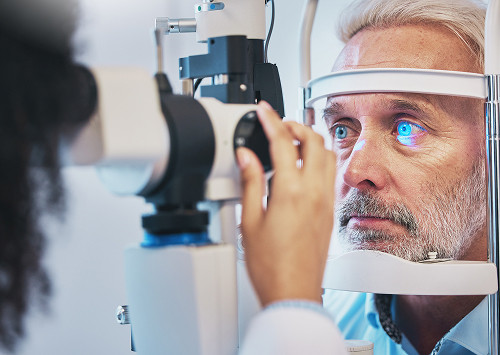
Before the procedure, the doctor examines the cornea and orders corneal tests. Corneal tomography, topography, and pachymetry are commonly used: they help assess the shape, curvature, thickness, and areas of maximum weakening.
We determine the recent history of vision changes, including any surgeries, injuries, inflammation, allergies, or frequent eye rubbing. This information helps us understand the cause of the changes and the speed of the process.
Crosslinking Stages
The protocol depends on the condition of the cornea and the clinical objective. Typically, the patient undergoes preparation, local anesthesia with drops, surface preparation, tissue saturation with a solution, and light exposure.
The main stages are as follows:
- The doctor reviews the examination data again and clarifies the plan.
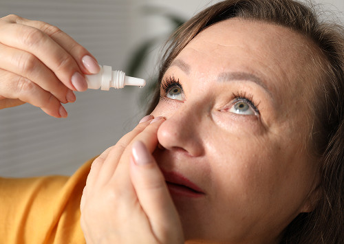
- Anesthetic drops are instilled into the eye.
- The corneal surface is prepared according to the selected protocol.
- Riboflavin is applied to the cornea until the tissue is sufficiently saturated.
- Then ultraviolet light is applied in a controlled manner.
- After completion, the doctor gives recommendations and schedules follow-up examinations.
We explain in advance how long the visit will take and what sensations are expected afterwards.
Anesthesia and patient sensations.
During the procedure, drop anesthesia is used. There is usually no significant pain, but the patient may feel light, pressure from the eyelid retractor, and tearing. After the drops wear off, burning, photophobia, and blurred vision may occur.
The doctor will warn you in advance about possible symptoms, how to use the prescribed medications, and when to contact the clinic. You cannot change the regimen on your own.

Frequently Asked Questions
Patients typically ask about pain, timeframes, prognosis, and risks. We answer questions directly, without making general promises.
Is crosslinking painful?
How long does recovery take?
Can vision be improved after the procedure?
Are there any contraindications?
Sign up for a consultation

If you already have a diagnosis or your doctor has told you that your cornea has become thinner, it's best not to delay an in-person consultation. The sooner you understand the progress, the more accurately you can choose your treatment plan.
What we recommend next
Schedule an ophthalmologist consultation at K+31 and bring your previous examination results, if available.
We will conduct a diagnosis, assess the risks, explain whether corneal crosslinking is necessary, and choose a safe route:
- Consultation
- Examination
- Procedure
- Post-treatment follow-up
If surgery is indicated, we will discuss preparation, sensations, limitations, and follow-up time in advance.
References
- https://cr.minzdrav.gov.ru/ — Clinical guidelines of the Russian Ministry of Health
- https://www.mediasphera.ru/journals/vestnik-oftalmologii — A specialized peer-reviewed journal
- https://www.rmj.ru/ — A Russian-language medical journal
- https://eyepress.ru/ — Russian-language ophthalmology resource
- https://cyberleninka.ru/ — scientific articles in Russian
- https://www.elibrary.ru/ — database of peer-reviewed publications in Russian
Our doctors


This award is given to clinics with the highest ratings according to user ratings, a large number of requests from this site, and in the absence of critical violations.

This award is given to clinics with the highest ratings according to user ratings. It means that the place is known, loved, and definitely worth visiting.

The ProDoctors portal collected 500 thousand reviews, compiled a rating of doctors based on them and awarded the best. We are proud that our doctors are among those awarded.
Make an appointment at a convenient time on the nearest date
Price
Other services

Appointment to the doctor
Reviews


Our clinics

Application “Personal Account K+31”


What is corneal collagen crosslinking?
Crosslinking is an ophthalmological procedure that increases the strength of the corneal stroma. The stroma is the main transparent layer; its condition determines the shape of the cornea and the quality of light transmission. This method is used when the tissue becomes thinner, weaker, and gradually changes shape.
How does the CXL method work?
The method is based on the reaction between the drug riboflavin and controlled exposure to light. A solution is applied to the cornea, then ultraviolet radiation is applied at predetermined parameters. Additional bonds are formed between collagen fibers in the tissue, making the cornea more resistant to stretching. The international name for this method is cross-linking.
This stabilizes the cornea. Potential vision improvement is discussed by the doctor only after assessing the shape, thickness, and optical parameters.
How does crosslinking differ from other treatment methods?
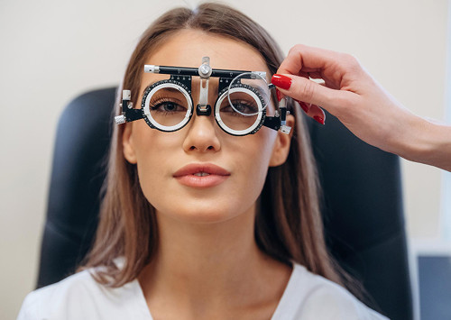
Glasses and contact lenses help improve vision, but they don't strengthen the cornea. Intrastromal segments change its geometry, and a corneal transplant is needed for severe changes. Corneal crosslinking improves tissue strength and helps slow further weakening.
Therefore, keratoconus treatment is often staged. First, the doctor checks the progress, then decides whether monitoring and correction are sufficient or whether a strengthening procedure is needed.