
Aphakia (absence of a natural lens): causes, symptoms, and vision correction
When the natural lens is missing from the eye, vision becomes blurred. Patients have difficulty reading and distinguishing details, have poor tolerance for bright light, and experience difficulty quickly defocusing.
At K+31, we begin with diagnosis and treatment selection. Aphakia requires accurate diagnosis, but in most cases, vision can be significantly improved.

specialists

equipment

treatment







How aphakia affects vision and well-being

An eye without a lens becomes highly farsighted. The person can see light and large outlines, but details are blurred.
Decreased Visual Acuity
Visual acuity usually decreases noticeably. In unilateral cases, the other eye partially helps. Anisometropia—a significant difference in optical quality between the eyes—is possible.
Image Distortion and Photophobia
Symptoms of aphakia can include glare, halos, changes in the size of objects, and discomfort in bright light. Photophobia is aggravated by inflammation or clouding of the ocular media. These complaints should be examined in person.
Double vision, rapid visual fatigue, and difficulty focusing
Double vision may occur when the two eyes function differently. Headaches may occur after reading, and a sensation of an unstable image may develop. Near focusing is impaired due to loss of accommodation. These symptoms of aphakia are especially significant after surgery or injury.
How do we diagnose aphakia?
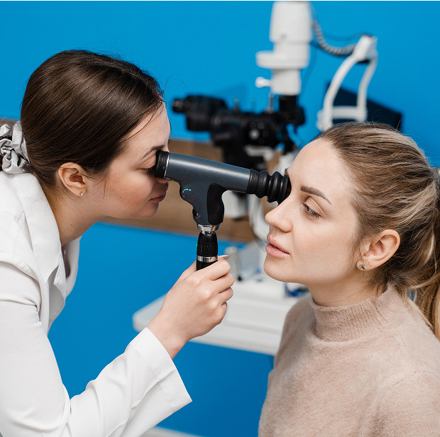
The diagnosis of aphakia begins with a consultation and examination. We determine when the vision changed and whether there was a cataract, injury, or surgery. Then we assess visual potential.
Visual Acuity and Refractive Sight Test
First, we check the visual acuity without glasses or contact lenses. Then, we select a trial correction and observe how much sharper the image becomes.
Next, we evaluate refraction—the optical parameters of the eye. If aphakia affects one eye, we separately check whether the patient comfortably tolerates the difference between the two eyes.
Biomicroscopy and Fundus Examination
Biomicroscopy allows us to examine the anterior segment of the eye under magnification. We examine the appearance of the cornea, iris, and pupil, and look for any remnants of capsular structures and signs of inflammation. A fundus examination reveals whether the retina and optic nerve are intact.
Additional tests before selecting a correction
Before surgery, an ultrasound of the eye, optical biometry, keratometry, OCT, and intraocular pressure measurement may be necessary. Diagnosing aphakia helps choose a safe path.
Aphakia and pseudophakia: what's the difference?
These conditions are often confused. In both cases, the natural lens is no longer present. But the optical situation and subsequent management are different.
When the crystalline lens is missing
Absence of a crystalline lens without an implant means the eye requires external or surgical refractive compensation. We evaluate the second eye, lens tolerance, the anterior segment, and the fundus.
When an artificial lens is installed
Pseudophakia means that an artificial lens is installed in the eye. It takes over the optical function of the natural lens. Vision quality depends on:
- Lens calculation
- Transparency of the media
- Optic nerve condition
Why is it important not to confuse these conditions?
If the absence of a crystalline lens is confirmed, one approach is required. In the case of pseudophakia, the examination is structured differently. A misunderstanding of terminology can lead to inconvenient optics and false expectations.
General information
Modern Methods of Aphakia Correction
Aphakia correction can be optical or surgical. We discuss clarity, care, limitations, and risks. You can't choose strong lenses on your own.
Main options:
| Method | When suitable | Advantages | Limitations |
|---|---|---|---|
| Glasses | If glasses are possible | Fast, without intervention | Distortions are possible |
| Contact lenses | If well tolerated | More physiological optics | Care and Regime |
| IOL / Intraocular Lens | Based on indications | Closer to natural optics | There are contraindications |
The table helps you understand the direction. The choice depends on the examination and visual tasks.
Glasses
Glasses may be suitable for bilateral aphakia or as a temporary solution. With high plus power, lenses can change the image size. Therefore, tolerance is checked in advance.
Contact lenses
Contact lenses are located closer to the eye, so they often provide a more natural optics. They can be convenient for unilateral aphakia. Before prescribing, we evaluate the ocular surface and the risk of inflammation.
Secondary IOL/Artificial Lens Implantation
Secondary IOL implantation is considered if an internal lens has not been previously implanted or external correction is poorly tolerated. The doctor evaluates:
- Capsular support
- Iris
- Anterior chamber
- Fundus
The IOL is selected based on calculations, and the intraocular lens is fixed only when sufficient conditions for safety are met.
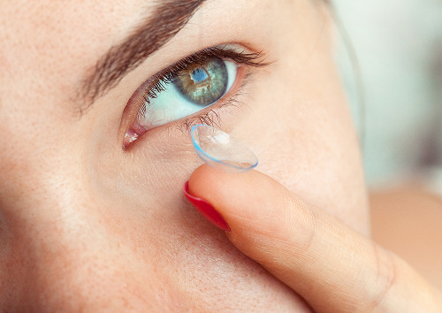

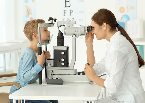

A scheduled ophthalmologist consultation is necessary if you experience persistent blurring after surgery, suspected lens displacement, intolerance to optics, or a significant difference in vision between your eyes.
You should see an ophthalmologist immediately if you experience pain, trauma, flashes, a dark "curtain," severe redness, or a sudden decrease in vision.
If your vision has decreased sharply or you experience flashes and pain, don't wait - see an ophthalmologist as soon as possible.
If aphakia is suspected, pay attention to the following signs:
- Sudden decrease in vision
- Feeling of blurriness and distortion
- Double vision
- Photophobia
- Easy eye fatigue
- Difficulty reading and Focusing
After such complaints, you shouldn't buy strong lenses yourself. Similar complaints can occur with inflammation and damage to the fundus.
Frequently Asked Questions
What is aphakia in simple terms?
Can vision be restored with aphakia?
What is best for aphakia: glasses, contacts, or an artificial lens?
How does aphakia differ from pseudophakia?
In aphakia, the natural lens is absent. In pseudophakia, the intraocular lens is already in place, taking over the optical function. Therefore, monitoring is structured differently.
If vision remains blurred postoperatively or following an injury, it is better to undergo an in-person assessment.
At K+31, we conduct an examination, explain the results in understandable language, and select a treatment plan without unnecessary treatment steps.
Treatment for aphakia is structured as a sequential process: diagnosis, decision-making, monitoring, and follow-up.
References
- MSD Manual — Aphakia (Russian version): https://www.msdmanuals.com/ru/home/eye-disorders/lens-disorders/aphakia
- MSD Manual — Aphakia for Professionals (Russian version): https://www.msdmanuals.com/ru/professional/eye-disorders/lens-disorders/aphakia
- Official Clinical Guidelines Portal Ministry of Health of the Russian Federation: https://cr.minzdrav.gov.ru/
- CyberLeninka — search by topic "aphakia": https://cyberleninka.ru/search?q=афакия
- CyberLeninka — search by topic "intraocular lens": https://cyberleninka.ru/search?q=интраокулная%20линза
- Russian Medical Journal — search by topic: https://www.rmj.ru/search/?q=афакия
Our doctors





















This award is given to clinics with the highest ratings according to user ratings, a large number of requests from this site, and in the absence of critical violations.

This award is given to clinics with the highest ratings according to user ratings. It means that the place is known, loved, and definitely worth visiting.

The ProDoctors portal collected 500 thousand reviews, compiled a rating of doctors based on them and awarded the best. We are proud that our doctors are among those awarded.
Make an appointment at a convenient time on the nearest date
Price
Other services

Appointment to the doctor
Reviews





















Our clinics


Application “Personal Account K+31”












Why does aphakia occur?
The cause affects the prognosis, the choice of optics, and the possibility of surgical intervention. We review the patient's surgical history, trauma, and previous examinations.
After cataract surgery
Long-term lens loss is most often associated with cataract surgery. After cataract surgery, an artificial lens is implanted. Sometimes implantation is delayed due to the condition of the capsule, inflammation, or traumatic changes. In such cases, aphakia correction is necessary.
After eye trauma
Trauma can damage the lens, displacing it, or causing its loss. The patient notices:
Here, treatment for aphakia begins with an assessment of the safety of the eye.
Congenital aphakia and rare cases
The congenital form is rare. In a child, the lens may be absent from birth or develop abnormally. In adults, rare causes are associated with lens displacement and previous interventions.