
Keratoglobus (corneal globe): symptoms, diagnosis and treatment
Keratoglobus is a rare corneal condition in which the cornea thins and becomes spherical. This reduces vision, causes distortion, and makes the cornea more vulnerable to injury. This condition is uncommon, but requires careful diagnosis and monitoring.
We'll explain how to recognize a corneal globus, what examinations are needed, and what methods can help preserve vision.

specialists

equipment

treatment






What is keratoglobus
The cornea is the transparent front layer of the eye, normally smooth and transmitting light. In this disease, it thins over almost its entire surface and bulges forward. As a result, light is refracted abnormally, and vision is impaired.
This condition is classified as an ectasia—a condition in which the cornea stretches and loses its strength. Due to diffuse thinning, it is less able to withstand stress. Therefore, it is important to make a prompt diagnosis and choose a safe approach. The earlier the thinning is detected, the greater the chance of preserving vision. Advanced cases are significantly more difficult to treat.
How does the shape of the cornea change with keratoglobus
With this disease, the cornea thins uniformly across its entire surface. Because of this, it takes on a rounded, spherical shape. This surface refracts light unevenly, resulting in blurred and distorted vision. Regular glasses are of little help, as the shape is far from perfect. Therefore, finding the right correction is more difficult than with myopia.
With keratoglobus, the entire optical system of the eye changes. Light rays pass through the cornea at different angles, so the image on the retina is not accurately collected. This results in:
- Blurred edges
- Halos around light sources
- Difficulty reading
- Emergent eye fatigue
The more severe the thinning, the more difficult it is to find the right glasses. The doctor evaluates the shape of the cornea based on the examination data and selects a correction that will provide the clearest and safest vision.
What is the difference between keratoglobus and keratoconus?
In keratoconus, the cornea bulges outward, forming a cone in one area, and the changes are usually localized. In keratoglobus, the thinning covers a larger area, and the cornea becomes spherical. Because of this, the cornea with this type of thinning is usually more vulnerable to injury.
| Signature | Keratoglobus | Keratoconus |
|---|---|---|
| Corneal shape | Diffuse spherical thinning | Conical bulge |
| Area of change | Often more extensive | Usually more localized |
| Risk | Severe corneal vulnerability | Progressive astigmatism |
| Approach | Individual correction and monitoring | Correction, stabilization, monitoring |
Why does keratoglobus develop?

The causes of this rare disease are not fully understood, but the main factors are known. Some cases are associated with congenital characteristics, while others are due to other conditions. Often, several factors are at play simultaneously. Let's break them down into groups.
Congenital characteristics and hereditary factors
The disease is often associated with a congenital weakness of the connective tissue. In such cases, the disease manifests early and may be associated with other connective tissue conditions. Heredity plays a significant role here.
Therefore, in cases of family history, early examinations are recommended for children. This helps to detect changes early.
If a hereditary form is suspected, the doctor will ask:
- Have any relatives had similar diagnoses?
- At what age did the complaints begin?
- How has vision changed?
This information helps assess the risk of progression and determine how often a child or adult patient needs follow-up examinations and repeat corneal diagnostics.
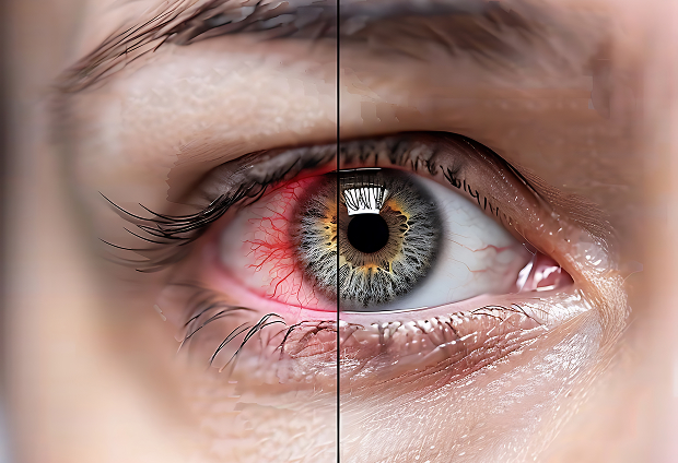
Concomitant conditions and risk factors
Sometimes, thinning of the iris develops as a result of general connective tissue diseases. Chronic inflammation and metabolic disorders also contribute.
Therefore, when making a diagnosis, the doctor takes into account the overall health of the body and concomitant diseases. This helps to understand the cause of the thinning and plan appropriate care.
Trauma and inflammatory processes
Past injuries and prolonged inflammation can weaken the iris. These are especially dangerous against the background of an already thinned iris. Therefore, it is important for these patients to protect their eyes from injury. Avoid rubbing the eyes and avoiding potentially traumatic situations. Even minor impacts can damage the delicate iris.
General information
How do we diagnose keratoglobus
Diagnosing keratoglobus relies on an accurate assessment of the shape and thickness of the cornea. A simple examination is not enough. Let's review the main steps.
Ophthalmologist examination and slit lamp
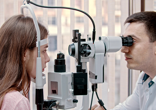
First, the ophthalmologist checks visual acuity and examines the cornea with a slit lamp. Under magnification, thinning and changes in the shape of the cornea are visible. The doctor asks how long the vision has been deteriorating and whether there have been any injuries.
During the examination, the corneal transparency, the condition of the tear film, the severity of astigmatism, and the eye's response to light are also assessed. These data help differentiate keratoglobus from other corneal ectasias and determine what further tests are needed.
The doctor also discusses the patient's complaints: how has vision changed, whether glasses are helping, whether there is double vision, glare, or rapid eye fatigue. This discussion is important for choosing a treatment plan, as the instrumental data must be compared with the patient's actual visual needs.
Pachymetry, corneal topography, and tomography
The corneal thickness is measured using pachymetry, and the surface shape is assessed using corneal topography. Additionally, a corneal tomography is performed, which shows its structure in detail. These data confirm the diagnosis and help monitor progress.
Anterior segment OCT and additional methods
For a detailed assessment, anterior segment OCT is used. This method shows the structure and areas of greatest thinning layer by layer. If necessary, other tests are prescribed to refine the treatment plan. Repeated examinations help determine whether the disease is stable or progressing. This determines treatment options.
FAQ
Short answers to the questions most frequently asked during appointments.
What is keratoglobus and how does it differ from keratoconus?
Is it possible to treat keratoglobus without surgery?
What tests are needed for diagnosis?
When should you urgently see a doctor?

The Registry of Clinical Guidelines of the Ministry of Health of the Russian Federation — https://cr.minzdrav.gov.ru/
The Russian Society of Ophthalmologists — https://www.ophthalmology.ru/
The Bulletin of Ophthalmology (Mediasphera) — https://www.mediasphera.ru/journal/vestnik-oftalmologii
The Russian Medical Journal — https://www.rmj.ru/
CyberLeninka - search on the topic of keratoconus - https://cyberleninka.ru/
eLIBRARY - publications on the topic of keratoconus - https://elibrary.ru/
CyberLeninka - search on the topic of keratoglobus - https://cyberleninka.ru/
Our doctors





















This award is given to clinics with the highest ratings according to user ratings, a large number of requests from this site, and in the absence of critical violations.

This award is given to clinics with the highest ratings according to user ratings. It means that the place is known, loved, and definitely worth visiting.

The ProDoctors portal collected 500 thousand reviews, compiled a rating of doctors based on them and awarded the best. We are proud that our doctors are among those awarded.
Make an appointment at a convenient time on the nearest date
Price
Other services

Appointment to the doctor
Reviews





















Our clinics


Application “Personal Account K+31”












Symptoms of keratoglobus
The symptoms of keratoglobus are similar to those of other corneal diseases, so they are easy to underestimate. They worsen as the cornea thins. Let's look at the main ones.
Vision loss and image distortion
The main symptom is decreased vision, which is poorly corrected by regular glasses. Straight lines appear curved, and images become blurry. The person notices that their vision is deteriorating, and correction is less and less effective. Often, vision differs in both eyes. This is also a reason to undergo an examination.
Astigmatism, photophobia, and rapid eye fatigue
Due to the uneven surface, severe astigmatism develops. This is accompanied by photophobia and rapid eye fatigue. These complaints intensify with visual strain and in the evening. Long periods of time spent working at a screen are more difficult to tolerate.
When to See a Doctor Urgently
Urgent care is needed if vision has deteriorated sharply, or if pain, photophobia, or redness develops. Symptoms following an eye injury are especially dangerous, as the thinned cornea is more easily damaged. Schedule an appointment for a diagnosis if you notice the following signs:
If blurred vision or pain develops after an eye injury, the patient needs an immediate consultation.