Optical examination of the posterior segment of the eye in Moscow
An optical examination of the posterior segment of the eye helps evaluate the structures responsible for central and peripheral vision. The doctor primarily examines the retina, macular area, vitreous, and optic nerve.
We perform diagnostics using modern equipment, explain the results to the patient in understandable language, and immediately determine the next steps.

specialists

equipment

treatment


When is the test prescribed?

Symptoms that require an eye exam
Symptoms that require an eye exam:
- Floaters before the eyes
- Flashes of light
- Blurred vision
- Distorted lines
- Sudden decrease in vision
- Appearance of a dark spot in the center of the image
- Feeling of a curtain or loss of a portion of the visual field
If vision has suddenly decreased, urgent medical attention is needed. In this situation, it is best not to postpone a scheduled eye exam, as time can affect the prognosis for some conditions.
What diseases and changes can this method detect?
A posterior eye exam helps the doctor detect signs of vascular, degenerative, inflammatory, and traction changes. Common reasons for examination include diabetic retinopathy, age-related macular degeneration, suspected retinal tears, hemorrhages, macular edema, vitreoretinal changes, and retinal detachment.
Retinal OCT is particularly useful for assessing tissue thickness, fluid presence, and the condition of the photoreceptor layers and macular area.
General information
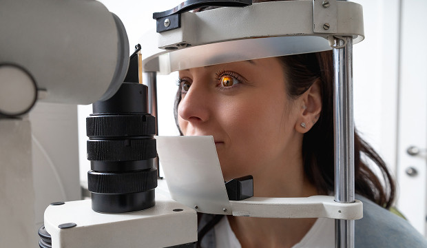
How is an optical examination of the posterior segment of the eye performed?
Is any preparation necessary before the procedure?
We usually only ask that you remove your contact lenses before the examination. Sometimes mydriasis—pupil dilation—is necessary for a better view of the posterior segment.
If pupil dilation drops are used, it is important for the patient to be aware of a temporary decrease in focus and increased sensitivity to light. It is recommended not to drive immediately after using such drops.
Examination stages: what the doctor does.
First, the doctor clarifies the patient's complaints, the duration of symptoms, the presence of injuries, surgeries, diabetes, hypertension, myopia, and hereditary factors. An ophthalmological examination is then performed, which helps determine which methods are needed for this particular patient.
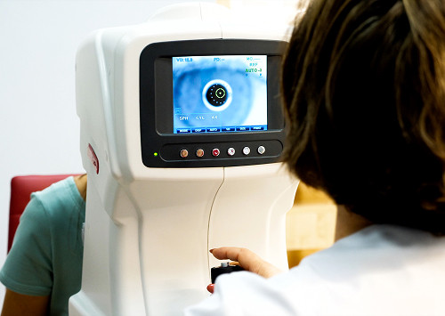
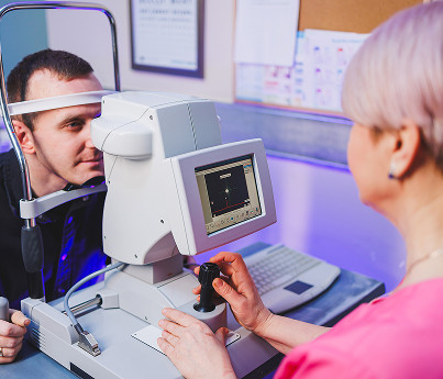
After the initial assessment, an OCT scan of the retina, a fundus examination, or other tests are performed as indicated. The doctor evaluates the image quality, assesses the macular area, the condition of the nerve, blood vessels, and vitreous, and then explains to the patient which changes require attention.
How long does the examination take and when are the results available?
The duration of the appointment depends on the scope of the diagnostic work. If the pupil needs to be dilated, the visit may be longer because the doctor needs to wait for the drops to take effect.
The patient receives the results on the same day of the appointment. We show the images, explain the key parameters, and prepare a doctor's report.


Frequently Asked Questions
What does a posterior segment optical examination reveal?
Do I need to prepare for the procedure?
Is a posterior segment optical examination painful?
How often should this examination be performed?
Book an appointment for a posterior segment optical examination

If you are concerned about flashes of light, floaters, blurred vision, image distortion, or your doctor has already recommended a retinal examination, schedule an appointment in Moscow. We will perform an OCT scan of the retina, evaluate the fundus, examine the posterior segment of the eye, and provide a clear report with further recommendations.
Scheduling an appointment is convenient for patients who want a quick examination, a doctor's report, and avoid wasting time on disjointed consultations. At our clinic, posterior segment examinations are performed with an emphasis on accuracy, safety, and a clear outcome for the patient.
Our doctors


This award is given to clinics with the highest ratings according to user ratings, a large number of requests from this site, and in the absence of critical violations.

This award is given to clinics with the highest ratings according to user ratings. It means that the place is known, loved, and definitely worth visiting.

The ProDoctors portal collected 500 thousand reviews, compiled a rating of doctors based on them and awarded the best. We are proud that our doctors are among those awarded.
Make an appointment at a convenient time on the nearest date
Price
Other services

Appointment to the doctor
Reviews


Our clinics

Application “Personal Account K+31”


What is a posterior segment optical examination?
An optical examination of the posterior segment of the eye is a comprehensive assessment of deep structures using modern imaging techniques. Depending on the patient's complaints and examination findings, the physician may use OCT, fundus examination, digital image capture, and additional methods if visibility is impaired.
Optical coherence tomography is a technology that creates a layered image of tissues and allows for the detection of changes that may be subtle during a routine examination.
What eye structures do we evaluate?
During the examination, we focus on the posterior segment of the eye because this is where the structures often affected by vascular, degenerative, and inflammatory processes are located.
The retina is responsible for image perception, and the macular area provides clear central vision, which is necessary for reading, computer work, and recognizing details. The optic nerve transmits information further, so its condition is important to check if glaucoma, neuro-ophthalmological disorders, and the consequences of vascular diseases are suspected.
How does this method differ from a routine examination?
The fundus may appear relatively normal, but edema, traction, thinning, or other initial abnormalities are already forming within the retina.
This is why posterior segment examination reduces the risk of missing early changes and helps determine whether the situation requires observation, treatment, or urgent referral to a specialist.