Transillumination (diaphanoscopy) of the eye in Moscow
The team of ophthalmologists at K31 Clinic offers its patients modern and time-tested examination technologies that help quickly and accurately diagnose and initiate treatment. One such technique is a specialized eye examination using a focused beam of light, known in medical practice as diaphanoscopy.
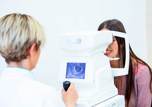
This procedure helps assess the transparency of the internal environment and identify various pathological lesions. Our clinic performs a comprehensive eye examination using cutting-edge equipment, so transillumination of the eye is performed in the most comfortable conditions possible.

specialists

equipment

treatment


When do we recommend diaphanoscopy of the eye?
Suspected Tumors and Cysts
We often use this method when patients are suspected of having various types of intraocular tumors. A dense tumor will block light rays, producing a distinct darkening of the pupil, while a hollow cyst filled with clear fluid will transmit almost all light. This allows the doctor to determine the density and structure of the identified lesion, which is crucial for developing a further treatment algorithm.
Unclear Complaints and Changes in the Anterior Segment
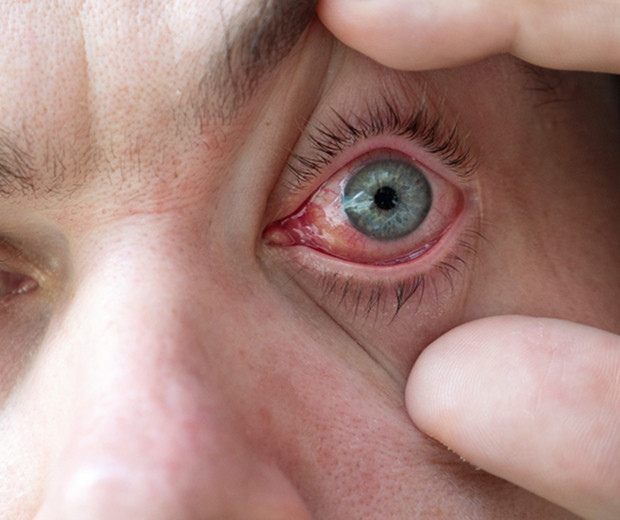
We recommend this examination if the patient has structural changes affecting the anterior segment of the eye. Focused light helps to examine in detail areas such as the iris and ciliary body, which are often involved in inflammatory processes or become the site of hidden degenerative lesions. This method helps detect small foreign bodies lodged in the deep layers of the sclera or embedded in the iris root area.
When is this method needed as part of a comprehensive diagnosis?
This eye diagnostic is prescribed at our clinic as an effective adjunct to a comprehensive examination.
When do we recommend transillumination of the eye:
- If tumors are suspected?
- If changes are observed in the anterior segment of the eye?
- If complaints are unclear and additional examination is needed?
- As part of a comprehensive examination by an ophthalmologist?
How is the examination carried out?
The examination process consists of several stages.
Is preparation necessary?
Ocular transillumination requires no special preparation: no diet or lifestyle changes are required. On the day of the procedure, women are advised to completely avoid wearing makeup on their face and eyes, as mascara or eyeshadow particles may create additional visual interference for the doctor during the examination.
Procedure Steps
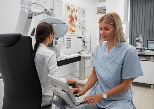
The examination is performed in a specially equipped room in a completely darkened room, which is necessary to achieve maximum contrast of the light beam. The patient is comfortably seated in a chair, after which drops are administered to dilate the pupil and local anesthesia is administered to completely eliminate discomfort when the device contacts the sclera.
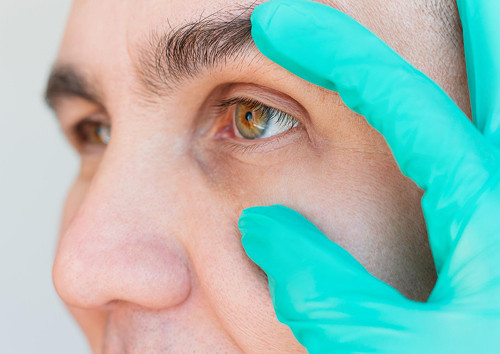
The doctor applies the tip of the illuminated device to the surface of the eyeball at various points, simultaneously assessing the pupil's luminescence through a special magnifying glass. The entire procedure takes just a few minutes and is extremely comfortable for patients.
When we communicate the results
The doctor announces the examination results immediately after the examination. Based on the data obtained, an official medical report is prepared and issued to the patient along with recommendations for further treatment or a referral for additional diagnostic procedures.
General information
Advantages of Transillumination in Our Practice
Fast and Comfortable for the Patient
In modern medical practice, speed of examination is of paramount importance. This procedure allows us to obtain important information about the internal structures of the visual organ in just one visit.
The patient experiences no discomfort, and exposure to the light beam lasts only a few minutes. This makes the method ideal for initial screening in complex clinical cases.
A Gentle and Safe Method
For many people, a safe diagnostic process is a key factor when choosing a clinic. The tissue transillumination device does not emit harmful radiation or exert aggressive thermal effects on delicate areas of the eye.
The procedure is painless, does not damage the integrity of the membranes, and can be repeated to monitor disease progression without any harm to the patient's health.
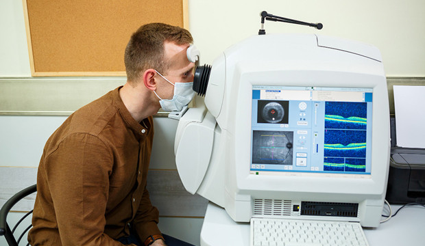
Complements slit lamp, ultrasound, and OCT
Each ophthalmological method has its own limitations, so we combine them to achieve the best results. Directional light transillumination does not replace other technologies, but rather serves as an important link between them. This method perfectly complements the data provided by a standard slit lamp.

Frequently Asked Questions
Is transillumination of the eye painful?
Is there any preparation needed for the procedure?
What does transillumination of the eye reveal?
Can ultrasound or OCT replace transillumination?
Book an appointment for transillumination (diaphanoscopy) of the eye in Moscow

If you're experiencing unexplained symptoms, deteriorating vision, or have been scheduled for a comprehensive eye examination, don't delay your visit to a specialist. Timely and accurate diagnostics help identify dangerous pathologies before they cause irreparable damage to your vision. At our clinic, you can undergo all necessary examinations quickly, efficiently, and in a comfortable, friendly atmosphere.
Schedule a consultation with an ophthalmologist in Moscow—we'll select the optimal diagnostic format, conduct a thorough examination, and answer all your questions. Our team of K31 doctors guarantees a personalized approach to each clinical case, the use of only certified equipment, and expert support throughout every stage of your eye care.
References
- Ministry of Health of the Russian Federation — official website: https://minzdrav.gov.ru/
- Register of Clinical Guidelines of the Ministry of Health of the Russian Federation: https://cr.minzdrav.gov.ru/
- Helmholtz National Medical Research Center of Eye Diseases: https://helmgoltz.ru/
- Scientific and Technical Complex "Eye Microsurgery" named after Academician S.N. Fedorova: https://www.mntk.ru/
- Ophthalmology Bulletin / Specialized Materials: https://www.eyepress.ru/
- Russian Medical Journal: https://www.rmj.ru/
Our doctors


This award is given to clinics with the highest ratings according to user ratings, a large number of requests from this site, and in the absence of critical violations.

This award is given to clinics with the highest ratings according to user ratings. It means that the place is known, loved, and definitely worth visiting.

The ProDoctors portal collected 500 thousand reviews, compiled a rating of doctors based on them and awarded the best. We are proud that our doctors are among those awarded.
Make an appointment at a convenient time on the nearest date
Price
Other services

Appointment to the doctor
Reviews


Our clinics

Application “Personal Account K+31”


What is transillumination (diaphanoscopy) of the eye?
This examination method is based on assessing the passage of light rays through various structures of the visual organ.
How the method works and what it allows us to see
A special instrument called a transilluminator is applied to the patient's sclera or eyelids, creating a powerful, focused glow. The ophthalmologist observes how this light penetrates the internal environment and evaluates the uniformity of the pupil's illumination.
Healthy tissue transmits light well, while any dense structures block it, creating a distinct shadow. This method allows for effective examination of the anterior segment of the eye, including the condition of the sclera and cornea, as well as assessing the deep internal zones for foreign bodies or areas of compaction.
How does transillumination differ from other diagnostic methods?
When performing standard transillumination, the specialist sees shadowy silhouettes of objects that are hidden from normal visual inspection. This method is indispensable in situations where standard optical media, such as the crystalline lens or vitreous body, have partially or completely lost their transparency due to a mature cataract or hemorrhage. In such circumstances, a normal examination becomes impossible, and transillumination helps obtain initial data on the condition of internal structures.