Gonioscopy in Moscow – examination of the anterior chamber angle for glaucoma
Gonioscopy in Moscow at the K+31 clinic helps the doctor see what a regular eye exam doesn't reveal: how open the anterior chamber angle is, how the aqueous humor outflow zone functions, and whether there are any signs important for diagnosing glaucoma. This procedure is especially important in cases of elevated intraocular pressure, suspected narrow angle, and the risk of the disease progressing without noticeable symptoms.

specialists

equipment

treatment


When we recommend gonioscopy
Specialists at the K+31 clinic recommend undergoing this diagnostic test in situations where it is necessary to clarify the condition of the drainage system and assess the risk of developing glaucoma.
It is recommended to seek help if there is:
- Suspected glaucoma
- Increased intraocular pressure
- Narrow or closing angle of the anterior chamber
- Monitoring after laser or surgical treatment
- Injuries, inflammations, and structural changes
Suspected glaucoma
If you have visual field changes, complaints of blurring, heaviness in your eyes, or a hereditary risk, your ophthalmologist may prescribe this procedure as part of a comprehensive glaucoma diagnosis. This procedure can determine the type of disease and assess the likelihood of fluid drainage blockage.
Increased intraocular pressure
High intraocular pressure does not always indicate glaucoma, but it requires an accurate assessment of the angle. By examining the angle, the doctor can determine how freely the aqueous humor circulates and whether there is a risk of developing the disease.
Monitoring after treatment, laser procedures or surgery
After surgeries and procedures, including laser treatment for glaucoma, it's important to monitor the condition of the structures. Gonioscopy in Moscow allows you to assess changes after the procedure and understand how effectively the fluid drainage system is working.
Changes after injuries and inflammation
Changes in the anterior chamber angle may appear after inflammation, injury, or previous ophthalmological diseases. Sometimes these changes affect fluid circulation and gradually increase intraocular pressure. In such cases, examination helps detect pathological changes early.
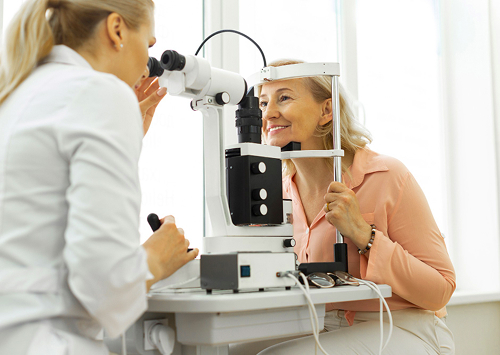
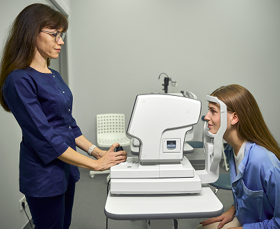
How gonioscopy is performed in our clinic
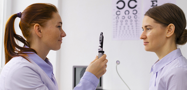
At the K+31 clinic, the procedure is performed on an outpatient basis and takes just a few minutes. An experienced ophthalmologist performs the entire procedure, explaining each step of the examination in detail to the patient.
Is any preparation necessary before the procedure?
No special preparation is usually required. Please inform the patient about any inflammations, allergies, previous surgeries, and medications you are taking. If you wear contact lenses, the doctor may ask you to temporarily remove them.
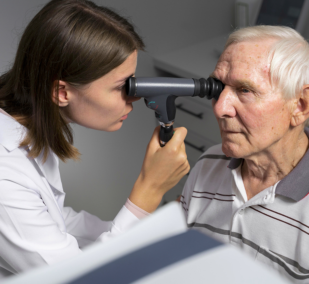
Stages of the examination
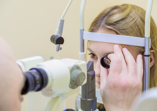
The patient is positioned in front of an instrument called a slit lamp. The doctor then uses a goniolens to examine the anterior chamber angle. Local anesthetic drops may be used for comfort.
During the procedure, the following is assessed:
- Width of the angle
- The condition of the structures between the iris and cornea
- The presence of changes in the fluid outflow zone
- The condition of the trabecular zone
There will be no pain. You will only feel a light touch of the lens.
How long does gonioscopy take and when does the patient receive the results?
The entire process takes a few minutes. The results are provided and explained immediately after the procedure. The doctor explains whether there are signs of pathology, how stable the intraocular pressure is, and whether additional vision diagnostics are needed.
General information
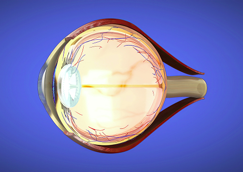
What does gonioscopy reveal?
The main goal of the procedure is to assess the condition of the structures responsible for the outflow of intraocular fluid.
Open or closed anterior chamber angle
The doctor determines the openness of the anterior chamber angle. This is the only way the ophthalmologist can detect angle-closure glaucoma, which develops rapidly and is accompanied by a sharp increase in pressure.
With open-angle glaucoma, the angle remains open, but the fluid filtration system is impaired. Such changes cannot be assessed without a special examination.
Condition of the trabecular meshwork and the drainage system of the eye
This type of diagnostic helps assess the condition of the area through which fluid outflow occurs. The specialist analyzes the appearance of the trabecular meshwork, looking for scars, adhesions, or other changes that impair the function of these structures.
This is an assessment of the eye's drainage system, which directly affects intraocular pressure.
How the results influence treatment strategy
The results help determine the appropriate treatment for the patient. Sometimes, observation and pressure monitoring are sufficient. In other cases, medication or surgery is required.
Accurate diagnosis of glaucoma is an opportunity to prevent vision loss and select treatment at an early stage.
"In our practice, gonioscopy often helps us see things that cannot be assessed without a special lens. We conduct the examination carefully, quickly, and immediately explain to the patient what the examination revealed and what steps are needed next," says an ophthalmologist at "K+31."
FAQ
What is gonioscopy?
Is gonioscopy painful?
Is preparation necessary?
How long does the procedure take, and when will the results be available?
Clinical Guidelines of the Ministry of Health of the Russian Federation — https://cr.minzdrav.gov.ru/
Ministry of Health of the Russian Federation — https://minzdrav.gov.ru/
Russian Medical Journal (Ophthalmology) — https://www.rmj.ru/
Bulletin of Ophthalmology — https://vestnik-oftalmologii.journals.eco-vector.com/
EyePress — Russian-language ophthalmology portal — https://eyepress.ru/
CyberLeninka — scientific publications — https://cyberleninka.ru/
Our doctors


This award is given to clinics with the highest ratings according to user ratings, a large number of requests from this site, and in the absence of critical violations.

This award is given to clinics with the highest ratings according to user ratings. It means that the place is known, loved, and definitely worth visiting.

The ProDoctors portal collected 500 thousand reviews, compiled a rating of doctors based on them and awarded the best. We are proud that our doctors are among those awarded.
Make an appointment at a convenient time on the nearest date
Price
Other services

Appointment to the doctor
Reviews


Our clinics

Application “Personal Account K+31”


What is gonioscopy and why do we prescribe this test?
Gonioscopy is a technique that allows an ophthalmologist to assess the area between the cornea and the iris, where the anterior chamber angle is located. This area contains the trabecular meshwork, part of the system through which aqueous humor drains from the eye. If this area is disrupted, intraocular pressure increases and damage to the optic nerve occurs.
The procedure uses a slit lamp and a special gonioscopy lens. The doctor sees structures that cannot be fully assessed during a standard eye examination. This is why this type of examination is considered an important part of modern glaucoma diagnostics.
This examination helps differentiate open-angle glaucoma from conditions associated with a narrow or closed angle. This is important for treatment selection, monitoring the condition, and assessing the risk of complications. Early diagnosis helps preserve vision and initiate glaucoma treatment before irreversible changes occur.