Secondary (Symptomatic) Glaucoma: Causes, Symptoms, and Treatment"

Secondary glaucoma is associated with a pre-existing problem: trauma, inflammation, surgery, vascular changes, or lens problems. Sometimes, it is caused by prolonged use of certain medications, especially without medical supervision. This causes increased intraocular pressure, placing strain on the optic nerve.

At "K+31," we examine the mechanism of the disease, assess vision risks, and recommend treatment after an in-person examination.

Best specialists
Best
specialists
Expert equipment
Expert
equipment
Advanced diagnostic treatment
Advanced diagnostic
treatment
Download the application “Personal Account K+31”
Everything about your health in one click!
Subscribe to social networks K+31
To keep up to date with current offers and receive useful advice about your health.

What is secondary glaucoma?

This is a group of conditions that disrupt the outflow of intraocular fluid or alter the pressure within the eye. This causes increased intraocular pressure, and the nerve fibers gradually lose their resistance to stress.

How does it differ from the primary form?

The primary form often develops without an obvious cause. With the secondary form, the doctor looks for a factor: eye trauma, uveitis, surgery, cataracts, vascular pathology, or long-term steroid drops. Symptomatic glaucoma requires pressure reduction and control of the underlying problem.

This difference affects the examination route and treatment strategy.

Symptom Secondary form Primary form
Cause Trauma, inflammation, surgery, or vascular disorder are present. There may be no obvious external cause.
Tactics Reduce blood pressure and treat the source of the disorder. Monitor blood pressure and nerve condition.
Dynamics May begin after a specific event Often develops gradually

This table provides guidance but does not replace an in-person consultation. The decision on treatment and its tactics depends on the condition of the cornea, lens, anterior chamber angle, retina, and optic nerve. This decision is made by an ophthalmologist.

Why is this disease dangerous for vision?

The main danger is related to damage to the nerve fibers. The optic nerve transmits images from the retina to the brain. And with prolonged pressure, some of the fibers stop working. Once the visual field narrows, some of the changes are difficult to reverse.

Causes and forms of secondary glaucoma
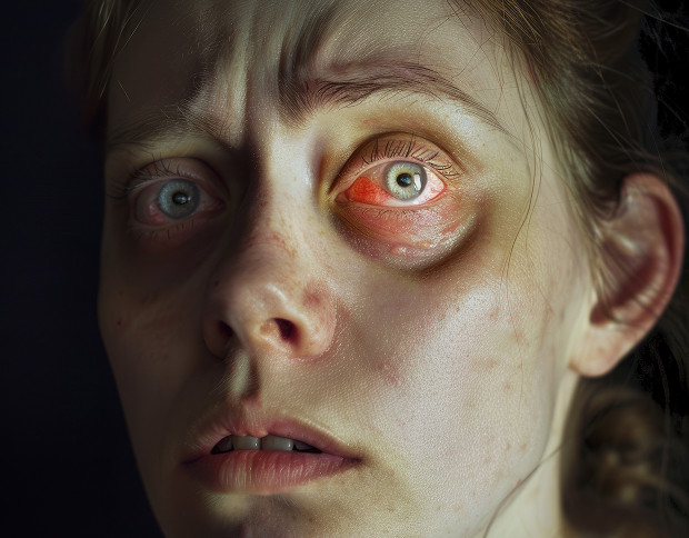
Secondary glaucoma always requires an investigation of the underlying cause.

After eye trauma

Post-traumatic glaucoma can occur after:

  • Blunt force
  • Wounds
  • Hemorrhage; with
    displacement of the lens

Pressure sometimes increases immediately, but in some patients the problem develops months later.

Due to inflammatory diseases

Inflammation within the eye interferes with normal fluid circulation. Inflammatory cells, protein components, pigment, and adhesions can block the drainage zone. In this situation, the ophthalmologist considers the activity of the process and the risk of relapse.

After eye surgery

Postoperative complications can alter the outflow of intraocular fluid. Causes include:

  • Inflammatory reaction
  • Hemorrhage
  • Scarring
  • Displacement of the artificial lens

Neovascular, phacolytic, steroid, and other forms

Neovascular glaucoma is associated with abnormal vessels in the iris and anterior chamber angle. Phacolytic glaucoma occurs when the lens undergoes changes, causing protein deposits to promote inflammation and interfere with the normal outflow of aqueous humor. Regardless of the cause, symptomatic glaucoma requires a precise understanding of why intraocular pressure increases and which ocular structures are involved.

Symptoms of secondary glaucoma

These depend on the cause and rate of pressure increase. Some patients experience eye pain and redness, while others experience only fog or a feeling of pressure.

Early signs that are often ignored

Signs of concern include fogging, rainbow-colored halos around incandescent lights, intermittent pain, one-sided headache, and decreased vision. If these symptoms recur, an ophthalmologist should be consulted. Self-administering eye drops can worsen the problem.

When to see an ophthalmologist urgently

An urgent examination is necessary if there is a sudden deterioration in vision, pain, redness, nausea, rainbow-colored halos, severe photophobia, or trauma. The combination of pain and a rapid decrease in vision is especially dangerous. In such a situation, intraocular pressure can rise rapidly. In this case, using hormonal medications without a doctor's supervision is dangerous.

How vision changes with progression

As vision progresses, peripheral vision suffers. The person:

  • Has difficulty noticing objects to the side
  • May bump into doorways
  • May see spots or gaps

Visual fields can help identify losses that are difficult to describe in words.

Symptoms of secondary glaucoma

General information

Diagnosis of Secondary Glaucoma

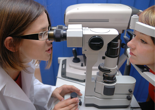
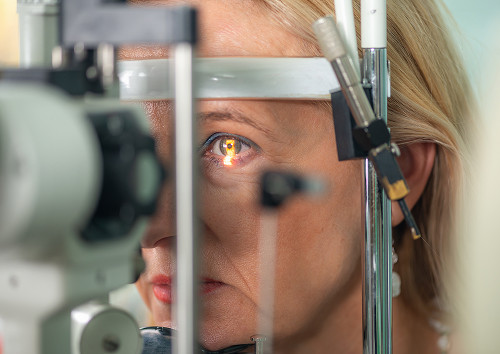
The examination begins with complaints, anamnesis, and physical examination. The doctor will review any injuries, surgeries, inflammations, vascular diseases, medications, and any deterioration. Intraocular pressure is then measured and the ocular structures are assessed.

Ophthalmologist Examination and Intraocular Pressure Measurement

During the examination, the ophthalmologist will check visual acuity, the cornea, iris, lens, anterior chamber, and fundus. Tonometry measures the pressure at the time of the examination. If fluctuations are suspected, the doctor will order a repeat pressure check or a 24-hour assessment.

Perimetry, OCT, Gonioscopy, Ophthalmoscopy

Perimetry evaluates the field of view and helps determine if there are any losses in peripheral vision. Tomography reveals the nerve fiber layer and the parameters of the optic nerve head. Examination of the anterior chamber angle helps understand the drainage pathways. Ophthalmoscopy is necessary to evaluate the fundus, retina, and optic nerve head. Therefore, the doctor may include several tests in the diagnosis, each of which clarifies a specific aspect of the picture:

  • Intraocular pressure measurement
  • Fundus examination
  • Visual field testing
  • Optic nerve OCT
  • Gonioscopy
  • Determining the cause of the disease

After the examination, the doctor explains the outcome to the patient: whether there are any signs of damage, how active the process is, and what steps are needed next. This analysis helps understand not only the test and imaging results but also the overall monitoring or treatment plan.

What tests help find the cause?

Sometimes an ultrasound of the eye, an anterior segment examination, a retinal examination, keratopachymetry, and a consultation with a general practitioner, rheumatologist, endocrinologist, or vascular specialist are needed. If vascular changes are present, the retina and signs of ischemia are assessed. Regarding the dosage form, the doctor clarifies what eye drops, injections, and systemic medications the patient is currently taking or has used in the past. Hormonal agents are especially important because they can increase intraocular pressure.
Diagnosis of Secondary Glaucoma

Treatment of Secondary Glaucoma

Treatment depends on the mechanism of increased pressure. The doctor considers:

  • Cause
  • Stage
  • Optic nerve condition
  • Drug tolerance
  • Ability to restore fluid drainage

The main goal is to maintain intraocular pressure at a safe level and preserve vision as much as possible.

Drug therapy

Drops can reduce the production of intraocular fluid or improve its drainage. The choice depends on the type of disease, inflammation, corneal condition, overall health, and drug tolerance. You should not stop taking prescribed medications on your own.

Laser Methods

Laser treatment is used for certain outflow obstruction mechanisms or retinal vascular changes. In some cases, the laser helps improve fluid circulation. In others, it reduces the stimulus for pathological vessel growth. The method is selected after an examination.

Surgical Treatment

This is considered when pressure cannot be maintained at a safe level or there is an anatomical cause that drops cannot address. The doctor can discuss:

  • Antiglaucoma surgery
  • Drainage systems
  • Crystal lens surgery
  • Combination treatment strategies

The extent of surgery depends on the damaged structures.

Treatment of the underlying disease is the key to success

Treatment of secondary glaucoma is more effective when the doctor controls the underlying cause. In cases of inflammation, it is important to reduce the activity of the process; in cases of vascular pathology, it is important to evaluate the retina and the overall treatment; and in cases of medication, it is important to adjust the medication. Therefore, an individual treatment plan is developed after a full assessment.

Treatment of Secondary Glaucoma

How We Approach Secondary Glaucoma Treatment

At "K+31," we guide the patient along a path:

  • Find the cause
  • Reduce pressure
  • Protect the optic nerve
  • Then check the results

We explain what changes are already present and what indicators we will monitor. Symptomatic glaucoma requires consistent management.

Individualized plan tailored to the cause of the disease

The individualized treatment plan includes examinations, therapy, monitoring schedules, and signs for unscheduled visits. The doctor considers the cause of the increased pressure in the eye: inflammation, vascular changes, or the consequences of trauma.

The focus of treatment and the frequency of follow-up depend on the cause. The patient receives an explanation of why this particular treatment plan has been chosen and how it helps preserve vision.

Monitoring Intraocular Pressure Dynamics

A single normal measurement does not always confirm stability. We evaluate intraocular pressure along with the patient's complaints, optic disc, corneal thickness, and functional tests. Repeated tonometry and pressure monitoring help determine whether therapy is adequate.

Monitoring and Adjusting Therapy

After improvement, monitoring continues. It is important for the doctor to ensure that visual function is stable and that the condition is not progressing. Regular vision monitoring and prevention of complications help detect deterioration early.

How We Approach Secondary Glaucoma Treatment

Why it's important not to delay treatment

Secondary glaucoma is dangerous because the underlying cause can continue to worsen the condition. Pressure sometimes drops temporarily, then rises again:

  • Active inflammation
  • Vascular problem
  • Medication reactions

If you experience pain, redness, fog, injury, or a sudden decrease in vision, it's best to get examined immediately.

Risks to the optic nerve

Long-term pressure damages the optic nerve and increases the risk of blindness. The degree of risk depends on the level of pressure, duration of the process, blood supply, age, and underlying medical conditions.

The doctor's goal is to stop or slow the damage while visual function remains functional.

What can be saved with early treatment?

Early treatment helps preserve vision that has not yet been lost. The doctor can reduce pressure, eliminate the triggering factor, prescribe observation, and promptly adjust treatment. The prognosis depends on:

  • Stage
  • Causes
  • General condition of the eye
Why it's important not to delay treatment

Our Advantages

At K+31, patients undergo an examination and receive an explanation of the results in a single process. We don't limit ourselves to pressure readings: for secondary cases, it's important to understand the source of the disorder.

Comprehensive Diagnostics

A comprehensive assessment includes an examination, pressure, nerve condition, anterior chamber angle, retina, and associated factors. If necessary, we involve additional tests and related specialists.

The patient receives a medical report that helps determine treatment and follow-up schedules.

Focus on the cause, not just the symptoms

If only complaints are addressed, the disease can continue to damage the eye. Therefore, we look for the factor causing increased intraocular pressure and assess its activity. In this case, treatment tactics become more precise.

A clear treatment plan and patient support

After the appointment, the patient knows which medications to use, when to return for a follow-up, and which symptoms require urgent contact with the doctor. If necessary, we discuss laser treatment or surgery, but strictly according to the indications. The main goal of support is to preserve vision and respond promptly to changes.

Our Advantages

Frequently Asked Questions

Can secondary glaucoma be completely cured?

Sometimes, after eliminating the underlying cause, pressure can be stabilized for a long time. However, secondary glaucoma may have already damaged the nerve fibers, and such changes are not fully restored. The answer depends on the type, stage, and speed of treatment.

Is secondary glaucoma always accompanied by pain?

No, pain is not always present. Symptoms of secondary glaucoma may include fog, decreased clarity, rainbow-colored rings, a feeling of pressure, redness, or a gradual narrowing of vision. Therefore, symptomatic glaucoma after injury, inflammation, and surgery requires monitoring.

Can vision be restored if it has already decreased?

Part of the loss may be due to swelling, inflammation, cataracts, or high pressure, in which case improvement is possible after treatment. The nerve damaged by glaucoma usually does not fully recover. The earlier secondary glaucoma is diagnosed, the higher the chance of preserving useful functions.

Do I need to undergo regular follow-up after treatment?

Yes, even if I feel well, I need to be monitored. Blood pressure can rise again if inflammation reactivates, the vascular situation changes, or a new reaction to medications occurs. Treatment of secondary glaucoma often requires ongoing monitoring. Therefore, at K+31, we create a clear and understandable schedule of appointments for patients.

References

Our doctors

Karapetyan Arevik Samvelovna
Experience 12 years
Make an appointment
Karapetyan
Arevik Samvelovna
Ophthalmologist, Laser Surgeon
Batalina Larisa Vladimirovna
Experience 33 years
Make an appointment
Batalina
Larisa Vladimirovna
Deputy chief physician for ophthalmology, ophthalmic surgeon, leading specialist in laser vision correction
Amiryan Anush Gamletovna
Experience 28 years
Make an appointment
Amiryan
Anush Gamletovna
Ophthalmic oncologist surgeon
Medvedev Yulia Alexandrovna
Experience 11 years
Make an appointment
Medvedev
Yulia Alexandrovna
Ophthalmologist, Laser Surgeon
Rudkovskaya Elena Mikhailovna
Experience 38 years
Make an appointment
Rudkovskaya
Elena Mikhailovna
Ophthalmologist
Tegniryadnova Ekaterina Valerievna
Experience 20 years
Make an appointment
Tegniryadnova
Ekaterina Valerievna
Ophthalmologist, laser surgeon
Fadeeva Victoria Anatolievna
Experience 13 years
Make an appointment
Fadeeva
Victoria Anatolievna
Doctor ophthalmologist, laser surgeon
Shamsetdinova Leila Tagirovna
Experience 17 years
Make an appointment
Shamsetdinova
Leila Tagirovna
Pediatric ophthalmologist
Gavrilina Polina Dmitrievna
Experience 8 years
Make an appointment
Gavrilina
Polina Dmitrievna
Ophthalmologist, laser surgeon
Sugonyaeva Olga Yurievna
Experience 26 years
Make an appointment
Sugonyaeva
Olga Yurievna
Ophthalmologist, laser surgeon
Khlunovskaya Anna Nikolaevna
Experience 8 years
Make an appointment
Khlunovskaya
Anna Nikolaevna
Ophthalmologist
Akhiyarova Azalia Azatovna
Experience 4 years
Make an appointment
Akhiyarova
Azalia Azatovna
Ophthalmologist
Putintseva Polina Andreevna
Experience 6 years
Make an appointment
Putintseva
Polina Andreevna
Ophthalmologist
Agafonova Oksana Sergeevna
Experience 21 year
Make an appointment
Agafonova
Oksana Sergeevna
Ophthalmologist, pediatric
Kazennov Alexey Nikolaevich
Experience 19 years
Make an appointment
Kazennov
Alexey Nikolaevich
Ophthalmologist, ophthalmologist-surgeon
Petrova Maria Grigorievna
Experience 14 years
Make an appointment
Petrova
Maria Grigorievna
Ophthalmologist, ophthalmic surgeon
Mazmanyan Karen Akopovich
Experience 28 years
Make an appointment
Mazmanyan
Karen Akopovich
Ophthalmic surgeon
Suchkova Valeria Alekseevna
Experience 6 years
Make an appointment
Suchkova
Valeria Alekseevna
Ophthalmologist
Khubieva Salima Aslanbievna
Experience 7 years
Make an appointment
Khubieva
Salima Aslanbievna
Ophthalmologist, laser surgeon
Rytik Nina Petrovna
Experience 15 years
Make an appointment
Rytik
Nina Petrovna
Ophthalmologist
All specialists
2GIS Award
2GIS Award

This award is given to clinics with the highest ratings according to user ratings, a large number of requests from this site, and in the absence of critical violations.

«Good place» according to Yandex

This award is given to clinics with the highest ratings according to user ratings. It means that the place is known, loved, and definitely worth visiting.

Our doctors are laureates of the ProDoctors Award

The ProDoctors portal collected 500 thousand reviews, compiled a rating of doctors based on them and awarded the best. We are proud that our doctors are among those awarded.

Make an appointment at a convenient time on the nearest date

Price

Reception
Price
Initial Ophthalmologist Appointment
from 6 450 ₽
Repeat Ophthalmologist Appointment
from 5 700 ₽

Other services

Laser vision correction Phacoemulsification of cataract with IOL implantation of any complexity Vitreoretinal surgeries of any complexity (vitrectomy) Laser photocoagulation of the retina during tears and degenerative changes in the retina Laser photocoagulation for diabetic retinopathy Intravitreal administration of Anti-VEGF drugs, hormonal implants Antiglaucomatous operations using various drains YAG laser dissection of the posterior capsule for secondary cataract Cataract treatment Treatment of glaucoma Retinal dystrophic changes Vitreous pathology Injury to the eye Post-traumatic and postoperative complications Myopia treatment Farsightedness Treatment of astigmatism Retinal disinsertion Non-contact laser procedure SMARTSURFACE Keratoplasty Femto SUPER LASIK Laser coagulation of the retina of the eye treatment on Ellex Integre Pro Scan Treatment of the retina with the Navilas laser system Femtolaser Cataract Removal Selection of children's night lenses Laser vision correction Clear Офтальмолог-онколог в Москве Treatment of keratoconus Laser coagulation of the retina Refractometry Retrobulbar injections Restoration of vision Lacrimal ducts Eye wash Synoptophore Forbis ESOM Pediatric ophthalmologist Ophthalmologist Glasses Biomicroscopy Night lenses Photo of an eye Eyelid suturing Selection of lenses Retinal laser Vision test Vitrectomy Vitrectomy Vitrectomy Eye pressure Ophthalmoscopy Fundus Gonioscopy Discision Orbitotomy Ophthalmodiagnostics OCT of the optic disc Keratoplasty Crystalline lens replacement Lacrimal duct Eye biometrics Angle of strabismus Lacrimal point IOL implantation Heterophoria Refraction Needling pillows Sclerectomy Micropulse laser Eyelid massage Intravitreal injections Iridoplasty Accommodation Bacterial culture Ophthalmology Cyclophotocoagulation Laser trabeculoplasty Laser stimulation Makdel Iridotomy Goniopuncture Corneal crosslinking Perimetry Pachymetry Angio-OCT Tubular test Color vision Convergence test Autofluorescence Eye mobility Corneal diagnostics Corneal transplant Skiascopy Strabismus Injection in the eye Tonometry Schirmer's test Trabeculotomy Diaphanoscopy Foreign body Foreign body Eyelid neoplasms Retinal tear Pterygium Chalazion Pneumatic retinopexy Glaucoma Ultrasound of the eye Eye biometrics Improving vision Cataract removal An attack of glaucoma Night lenses Eyelid ptosis Vision correction Angiography of the eye Retinoblastoma Laser coagulation PRK Orbital exenteration Evisceration of the eye Exophthalmometry PresbyLasik Removal of the lens Retinal thrombosis Nystagmus Lacrimal duct Eye removal Optic nerve Eyelash removal Neoplasms of the eyelids Myopia PRP therapy Autorefractometry Anti-VEGF Iris melanoma Glaucoma treatment Megalocornea Macular degeneration AMVO-01 Visotronik Amblyopia Accommodation Ametropia Cataract Lagophthalmos Anisometropia Asthenopia The optic nerve Dry eye syndrome Strabismus Aphakia Retinal coagulation Eyesore Blepharitis Eyelid spasm Blepharochalasis Blepharophimosis Myopia Vitrectomy Presbyopia Glaucoma Cataract Conjunctivitis Secondary cataract Keratoconus Keratoglobus Keratitis
Make an appointment

Appointment to the doctor

Fill out the form, our managers will contact you within 15 minutes

Reviews

Everything is fine. She conducted a number of studies, listened attentively, was very pleasant in communication, explained everything clearly. I am satisfied.
21.07.2026
Svetlana S,
A great professional doctor, responsive, thoughtful, positive, attentive to the patient.
18.07.2026
Aleksandr K.
I signed up for a check-up and accidentally got to Azalea Azatovna and I am very happy that I got to her! He is a very sensitive and attentive doctor, the operation to restore vision was excellent and easy, for which thank you very much! After the operation, I realize that I shouldn't have waited and feared so much, with such a professional in his field, nothing is scary!thank you very much!!!
16.07.2026
Irina S,
The reception was professional and unobtrusive. Additional examinations and diagnostics were carried out using modern equipment. During the consultation and examination, Polina Dmitrievna explained everything in detail and intelligibly, gave me clear recommendations, and answered all questions in detail. Polina Dmitrievna is a polite, attentive and simply amazing doctor. The clinic as a whole enjoys the atmosphere, responsiveness of the staff and the professionalism of the specialists.
11.07.2026
Nikolay K.
Rudkovskaya E.M. is an excellent doctor, if you need an ophthalmologist, I go to her. A very competent specialist. Explains everything by diagnosis. Comprehensively evaluates the condition, checking all the parameters.
11.07.2026
Vera S.
Wonderful doctor!
10.07.2026
Natalya P.
An excellent specialist. I am very pleased with the reception. Thanks to the doctor
08.07.2026
Svetlana U.
I would like to express my sincere gratitude to all the staff of the K+31 clinic. Special thanks to the doctors of the ophthalmology department, Ekaterina V. Tegniryadnova, who has been seeing me for a long time, and Maria Grigorievna Petrova, who operated on me for a lens replacement. Everything went great! I would also like to mention the work of Diana's ophthalmology manager. I received comprehensive and timely answers to all my questions. THANK you so much to all of you!
Почему К+31?
К + 31 — full-cycle multidisciplinary medical centers, including the possibility of providing medical services of European quality level.
К + 31 — are leading doctors and diagnostics using high-tech equipment from world manufacturers (Karl Storz, Olympus, Siemens, Toshiba, Bausch&Lomb, Technolas, Zeiss, Topcon).
К + 31 — is ethical. The staff of K+31 clinics maintain open relationships with patients and partners. An individual approach to each patient is the basis of our service standards.
К + 31 — is modernity. On call 24/7: call center operators will answer your questions at any time and book you an appointment with doctors. Contact us by phone, through the feedback form on the website and Max.

Our clinics

Address K+31 Lobachevskogo 2

st. Lobachevskogo, 42/7

Contacts

+7 499 999-31-31

Opening hours

Mon-Fri: 08:00 – 21:00
Weekend: 09:00 – 19:00

Address K+31 on Lobachevskogo

st. Lobachevskogo, 42/4

Contacts

+7 499 999-31-31

Opening hours

Mon-Fri: 08:00 – 21:00
Weekend: 09:00 – 19:00

Address K+31 Petrovskie Vorota

1st Kolobovsky pereulok, 4

Contacts

+7 499 999-31-31

Opening hours

Mon-Fri: 08:00 – 21:00
Sat-Sun: 09:00 – 19:00

Address K+31 West

Orshanskaya, 16/2; Ak. Pavlova, 22

Contacts

+7 499 999-31-31

Opening hours

Mon-Fri: 08:00 – 21:00
Sat-Sun: 09:00 – 18:00
K+31 on Lobachevskogo
Didn't find the service you were looking for?

Экстренная помощь