
Blocked tear duct (tear duct obstruction): symptoms, diagnosis, and treatment
Tear duct obstruction most often manifests as constant tearing, where the tears seem to be stuck in the eye and overflowing. This causes discomfort, and when the tears stagnate, inflammation easily develops. Understanding the cause and choosing treatment is only possible after a doctor's examination. Let's look at how to recognize a blocked tear duct, its dangers, and methods for restoring drainage.

specialists

equipment

treatment






What is a blocked tear duct?
Normally, tears moisten the eye and drain through thin ducts into the nasal cavity. When an obstruction occurs, the outflow is disrupted, and fluid accumulates. This causes an obstruction, also known as a blockage.
The obstruction can be partial or complete, congenital or acquired. This determines the severity of the complaints and the treatment strategy. First, let's look at how the tear drainage system works.
How the lacrimal drainage system works
Tears form at the outer corner of the eye. They spread across the surface when you blink. They then collect at the inner corner. They then enter the puncta. The fluid then passes through the ducts into the lacrimal sac and drains into the nose through the nasolacrimal duct.
If any of these areas are narrowed or blocked, tear flow stops. The fluid stagnates, causing tearing. Stagnant conditions make infection more likely, so this problem should not be ignored.
How does obstruction differ from normal lacrimation?
The eye can water for various reasons: wind, allergies, irritation, or inflammation, such as conjunctivitis. In such situations, complaints are usually related to an external factor or infection. When drainage is obstructed, tears are constantly or almost constantly retained, usually on one side. This condition is called epiphora.
The lacrimal drainage pathways are responsible for tear drainage: the puncta, lacrimal canaliculi, lacrimal sac, and nasolacrimal duct. If a narrowing occurs in any area, the doctor may speak of dacryostenosis. Complete or severe blockage is called lacrimal duct obstruction.
It is difficult to distinguish between these conditions based on a single symptom. Therefore, if persistent tearing occurs, a lacrimal duct examination by an ophthalmologist is necessary. The doctor determines the specific location of the obstruction and decides whether the lacrimal duct can be restored conservatively or whether a procedure is required.
Symptoms that may indicate a problem

It's easy to suspect a drainage problem based on several characteristic signs. They increase gradually and often affect one eye. Let's take a closer look.
Constant tearing and lacrimation
The main symptom is chronic tearing, which is unaffected by wind or weather. Tears form at the edge of the eyelid and run down the cheek. Constant moisture irritates the skin around the eye.
Redness, swelling, and discharge
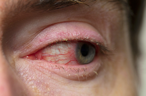
When congestion occurs, inflammation occurs, resulting in redness of the eye and swelling of the eyelid at the inner corner. Mucopurulent discharge often occurs, especially in the morning. If you press on the lacrimal sac, a cloudy discharge may emerge.
When is an urgent medical examination needed?
You should see a doctor immediately if you experience severe swelling, pain, and purulent discharge at the inner corner of the eye. This may be a sign of acute inflammation of the lacrimal sac. You should schedule a diagnostic appointment if you notice the following symptoms:
- Constant tearing
- Redness of the eye
- Swelling at the inner corner of the eye
- Mucopurulent discharge
- A feeling that tears "don't drain"
The sooner the cause is identified, the higher the chance of using gentle treatments and avoiding complications.
What is the danger of blocked tear ducts?
Impaired tear drainage can cause more than just discomfort. Stagnant tears can also create conditions for infection and chronic inflammation. Let's look at the main risks.
Risk of dacryocystitis and chronic inflammation
The most common complication is dacryocystitis, an inflammation of the lacrimal sac. It manifests as pain, swelling, and purulent discharge at the inner corner of the eye. Acute dacryocystitis requires prompt treatment, while chronic dacryocystitis is prone to recurrence.
A persistent source of infection near the eye is also dangerous for the eye itself. Therefore, treatment should not be delayed. The longer the infection persists, the higher the risk of complications.
Why you shouldn't delay diagnosis
At an early stage, the problem can often be resolved with gentle methods. When the inflammation becomes chronic, treatment becomes more complicated. Early diagnosis saves the patient time and energy.
How we conduct diagnostics

An examination helps determine the location of the obstruction and its severity. It usually takes one visit. Let's review the main steps.
Examination and patency check
The doctor examines the eyelids, lacrimal puncta, and lacrimal sac area. They assess for swelling and drainage when pressure is applied. This alone reveals a lot about the nature of the problem.
Lacrous duct irrigation
To check the drainage, the lacrimal ducts are irrigated. By checking whether fluid flows into the nose, the doctor determines the level and severity of the obstruction. This same procedure can sometimes help clarify the diagnosis.
Additional tests, if necessary
In complex cases, additional tests are prescribed to visualize the entire duct. A consultation with an ENT specialist is sometimes required. The scope of the examination depends on the specific situation.
What treatment and recovery methods do we use?
| Method | When used | What it provides | Characteristics |
|---|---|---|---|
| Irrigation | For initial symptoms and to clarify the diagnosis | Helps assess patency | Performed by a doctor |
| Probing | For severe or persistent obstruction | Restores tear outflow | Requires accurate diagnosis |
| Surgical restoration | For chronic obstruction and complications | Eliminates the cause Disorders | Individually selected |
Conservative therapy at an early stage
If inflammation is present, it is first relieved with topical agents as prescribed by a doctor. In children with congenital inflammation, a special massage of the lacrimal sac area can sometimes help. This approach is used under the supervision of a specialist.
Probing and irrigation
If the obstruction is persistent, probing of the lacrimal duct is performed to restore the lumen. The procedure is supplemented by irrigation to ensure patency. This minimally invasive treatment often helps without major surgery.
Surgical restoration of tear outflow
When gentle methods fail, surgery to restore the outflow is performed. The surgeon creates a new tear pathway around the obstruction. The method is selected individually based on the examination results.
In our practice, we see that the sooner a patient comes for a diagnosis, the higher the chance of resolving the problem with gentle methods – without prolonged inflammation and constant tearing. – ophthalmologist
General information
Why patients choose us
In this delicate area, accurate diagnostics and careful methods are essential. We structure our care so that patients quickly understand the cause and receive a solution. Below is what this means.
Comprehensive diagnostics without unnecessary procedures
We determine the level and cause of the obstruction in a single visit. We prescribe only the tests that are truly necessary. This saves the patient time and energy.
A gentle approach to adults and children
We work especially carefully with young patients. Any procedures in children are performed only according to medical indications and gently. We explain in detail to parents what is being done and why.
A clear treatment plan and support
We explain the diagnosis in simple terms and offer a clear path from diagnosis to restoration of tear flow. We support the patient until lasting results are achieved. If necessary, we involve related specialists.


FAQ
Short answers to the questions most frequently asked during appointments.
How do you know if your tear duct is blocked?
Is it possible to treat obstruction without surgery?
What are the dangers of a blocked tear duct?
When do irrigation or probing help?

Our patient journey is structured step-by-step: from examination to a clear plan. We'll explain what will happen at each stage.
Initial examination
The doctor will assess your complaints, examine your eyelids and puncta, and check your drainage. If necessary, perform an irrigation to determine the level of obstruction. The cause will become clearer during the appointment.
Treatment plan
Based on the examination, we'll suggest a treatment plan: from gentle procedures to surgery if indicated. We'll explain the advantages and limitations of each option. We'll make the decision together with the patient.
Recommendations for follow-up care
After treatment, we'll provide recommendations for care and monitoring. If you're prone to inflammation, we'll advise you on how to reduce the risk of flare-ups. If necessary, we'll schedule a follow-up visit.
Our doctors





















This award is given to clinics with the highest ratings according to user ratings, a large number of requests from this site, and in the absence of critical violations.

This award is given to clinics with the highest ratings according to user ratings. It means that the place is known, loved, and definitely worth visiting.

The ProDoctors portal collected 500 thousand reviews, compiled a rating of doctors based on them and awarded the best. We are proud that our doctors are among those awarded.
Make an appointment at a convenient time on the nearest date
Price
Other services

Appointment to the doctor
Reviews





















Our clinics


Application “Personal Account K+31”












Why does tear duct blockage occur?
There are several causes of drainage obstruction, and treatment depends on them. The obstruction may be present at birth or develop during life. Let's look at the main options.
Congenital causes
Congenital obstruction occurs in newborns when the duct has not fully opened at birth. The baby's eye constantly waters and becomes watery from the first weeks of life. In some cases, the duct opens on its own, but medical supervision is essential.
Inflammation, trauma, and scarring
In adults, the duct often narrows after inflammation and infection. Trauma to the face and nose, as well as scarring, damage the delicate tear ducts. Sometimes, obstruction occurs after surgery in this area.
Age-related changes and associated diseases
With age, the duct walls narrow, and drainage worsens. Chronic diseases of the nose and paranasal sinuses play a role. Therefore, if persistent tearing occurs, the doctor also evaluates the condition of the ENT organs.
Less commonly, tumors and systemic diseases are the cause. Such cases require a more comprehensive examination. The exact cause is determined by a specialist after an examination.