Retinal autofluorescence study: what the method shows
The retina changes before a person notices a decline in vision. There is no pain or complaints. However, areas may already appear in the fundus that require further diagnostic evaluation. Autofluorescence testing helps identify these areas without injections or eye contact.
At K+31, autofluorescence is prescribed when the doctor needs to more accurately assess the condition of the retina and determine further monitoring or treatment strategies.

specialists

equipment

treatment


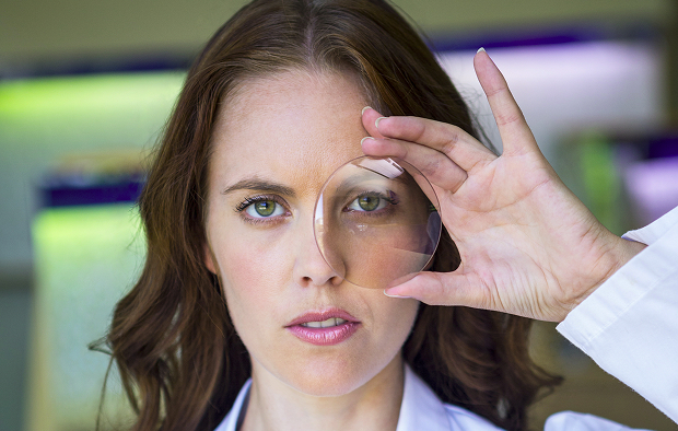
What is autofluorescence and why is it needed
Autofluorescence is the natural glow of individual structures in the fundus in response to light of a certain wavelength. The doctor evaluates not the glow itself, but its distribution in the image. Where the signal is smooth, where it is enhanced, and where it is reduced.
This method helps understand how tissue in the macula and other parts of the retina functions. It is useful when a routine examination is insufficient.
How the method works
The device directs light into the fundus and records the response signal. Most often, the doctor evaluates the retinal pigment epithelium—the layer of cells that supports photoreceptors and is involved in metabolic processes. If this layer is damaged, the signal changes.
Fundus autofluorescence does not require the injection of dye. It's a non-invasive method, so the examination is usually a relaxing experience: the patient looks at a marker, and the specialist takes images.
What exactly does the doctor evaluate?
A single light or dark area on an image doesn't explain anything. The doctor evaluates the appearance of the entire area: its shape, size, position relative to the macula, its relationship to the complaints, and the difference between the right and left eyes. Sometimes, comparing the two eyes helps determine the significance of the changes. When interpreting the image, several features are considered:
- Areas of increased or decreased signal
- Condition of the central retinal zone
- Consistency of data with the examination and OCT
The image provides the doctor with additional information. However, a final assessment is only possible after comparing all the data, because an image without a clinical picture can be misinterpreted.
In what cases is the study prescribed?
Autofluorescence testing is ordered when a more accurate assessment of the retina's condition is needed. This may be due to complaints or monitoring of a known condition. We do not prescribe it formally.
Suspected retinal changes
The patient may notice distortion of straight lines, a spot in front of the eye, decreased visual acuity, or difficulty reading. However, sometimes the opposite happens: there are no complaints, but during the examination, the doctor already notices areas that need to be examined more closely.
In such a situation, retinal diagnostics help determine whether the macula is affected, whether there are signs of atrophy, or metabolic disorders in the tissue. This makes it easier for the doctor to decide the next step: observation, additional tests, or treatment as indicated.
Monitoring existing diseases
This method is used for age-related changes in the macula, hereditary retinal diseases, the consequences of inflammation, vascular disorders, and changes associated with diabetes. Dynamics are important here.
The results can be compared with previous data. If the lesions are stable, the monitoring regimen is the same. If new areas appear, the strategy is revised.
Why is the examination important for early diagnosis?
Many processes in the fundus develop without pain or noticeable discomfort. A person attributes the decrease in clarity to fatigue, especially if the other eye sees well and shares the load. Therefore, early diagnosis is of great importance.
Retinal autofluorescence helps detect retinal changes that may not be obvious during a standard examination. This is especially important in cases of hereditary predisposition, diabetes, and age-related changes in the macula.
What the research results show
The image shows a signal map of the fundus. This allows the physician to understand where tissue appears stable and where there are signs of abnormalities. It is not recommended to evaluate the image independently.
Areas of increased and decreased autofluorescence
An increased signal may be associated with the accumulation of substances in cells or their increased load. A decreased signal sometimes corresponds to atrophy, cicatricial changes, or other causes. Interpretation depends on the diagnosis and the location of the area.
Fundus autofluorescence helps to identify not only the lesion but also its borders. During follow-up examinations, the doctor compares the images and monitors the dynamics.
What do changes in the pigment epithelium mean?
The retinal pigment epithelium supports the function of cells that perceive light. If this layer is damaged, vision may not deteriorate immediately.
The test results help suspect degenerative, hereditary, vascular, or inflammatory processes. However, a diagnosis is made only after comparing the data. The doctor considers the patient's complaints, visual acuity, macula, OCT, and physical examination.
Why is the result always assessed in a comprehensive manner?
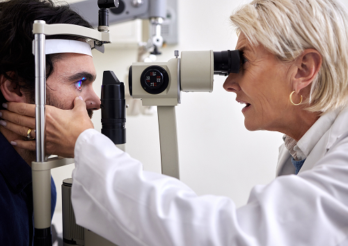
Diagnostics are necessary because different methods answer different questions. A fundus examination reveals the overall picture. OCT provides a layered image. And autofluorescence helps assess the condition of the pigment epithelium.
For the patient, the difference is easier to see in a table:
| Method | What it shows | Strength |
|---|---|---|
| Autofluorescence | Pigment epithelium, areas of signal change | Helps to detect early abnormalities |
| OCT | Layered structure of the retina | Shows the thickness and anatomy of tissues |
| Fundus photograph | General view of the retina and vessels | Records the image for Comparisons |
| Ophthalmologist examination | Complaints and eye condition | Links data to the clinical situation |
The scope of the examination is determined by the doctor. Sometimes a single image is sufficient, while other times a more extensive assessment is required.
General information
What diseases can this method help diagnose?
This test is used for various retinal conditions. It doesn't replace a doctor's diagnosis, but it helps identify important signs.
Age-related degenerative changes
With age, drusen, areas of thinning, and areas of atrophy may appear in the macular region. Initially, a person notices fatigue when reading or distorted lines. Sometimes, complaints appear later than changes in the images.
Retinal autofluorescence helps assess risk areas and compare them with OCT data. This is important for monitoring age-related macular degeneration.
Hereditary pathologies
With hereditary retinal diseases, changes can affect the center and periphery. The doctor evaluates the signal distribution. The symmetry of the two eyes and the relationship of findings to the complaints is also assessed.
An ophthalmological examination in such cases includes more than one test. We obtain a family history. Then we check visual function. Then we decide which methods are needed.
Vascular and metabolic disorders
In diabetic retinopathy, vascular disorders, and the consequences of inflammation, the retina suffers due to impaired tissue nutrition. The doctor examines whether the macula is affected. They also assess for signs of damage to the pigment epithelium.
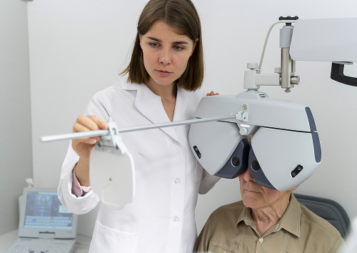
Retinal diagnostics for such conditions include OCT, pupillary dilation, fundus photography, and other methods as indicated. The range of tests is individualized.
FAQ
Before the examination, patients most often ask about safety, preparation, and the meaning of the results. These answers help guide the patient. The doctor makes the decision after the examination.
Do I need to prepare for the test?
Is autofluorescence safe?
Can this method replace other examinations?
When will the results be ready?

Russian Ministry of Health, Registry of Clinical Guidelines — https://cr.minzdrav.gov.ru/
CyberLeninka — Search on retinal autofluorescence — https://cyberleninka.ru/search?q=autofluorescence%20retina
CyberLeninka - search by fundus autofluorescence - https://cyberleninka.ru/search?q=фундус-аутфлюоресцентия
CyberLeninka — materials on changes in the retinal pigment epithelium — https://cyberleninka.ru/search?q=пигментный%20епителя%20ресчатки
CyberLeninka — age-related changes in the retina — https://cyberleninka.ru/search?q=возразная%20макулярная%20дегенерация%20аутфлюоресцентия
CyberLeninka — Diabetic retinopathy and autofluorescence — https://cyberleninka.ru/search?q=диабетическая%20ретинопация%20аутфлюоресцентия
eLIBRARY — Russian-language scientific publication database — https://elibrary.ru/
Our doctors


This award is given to clinics with the highest ratings according to user ratings, a large number of requests from this site, and in the absence of critical violations.

This award is given to clinics with the highest ratings according to user ratings. It means that the place is known, loved, and definitely worth visiting.

The ProDoctors portal collected 500 thousand reviews, compiled a rating of doctors based on them and awarded the best. We are proud that our doctors are among those awarded.
Make an appointment at a convenient time on the nearest date
Price
Other services

Appointment to the doctor
Reviews


Our clinics

Application “Personal Account K+31”


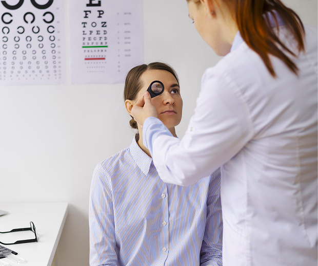
How is autofluorescence testing performed?
The examination is performed in the ophthalmology diagnostic room. The patient maintains a fixed gaze while the specialist takes images. There is no pain.
Is any preparation necessary?
No special preparation is usually necessary. There is no need to fast or stop taking regular medications. However, the doctor may dilate the pupil with drops if this will improve the image quality.
After dilating the pupil, blurred vision and sensitivity to light are possible. It is recommended to wear sunglasses and discuss driving.
How the procedure is performed step by step.
The specialist clarifies the patient's complaints. Then, the examination procedure is explained. The patient then rests their chin on the chin rest and looks at the indicated point. The standard procedure includes:
How long does the examination last and what does the patient feel?
The examination itself takes a few minutes. More time may be needed for pupil dilation and evaluation of the images. The patient is sensitive to light, may blink, and sometimes tires of focusing.
Autofluorescence is well tolerated by most patients. If you experience photophobia or lacrimation, please notify the specialist.