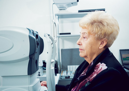
Partial Orbital Exenteration in Moscow

Tumorous lesions in the orbit cannot be assessed by their appearance. It is important to understand the extent of the lesion and whether healthy tissue can be preserved. Partial exenteration is performed when it is necessary to remove the affected structures without unnecessary expansion of the surgical procedure.

At "K+31," a request for orbital exenteration in Moscow guides the patient through an in-person examination, diagnosis, surgery, and follow-up.

Best specialists
Best
specialists
Expert equipment
Expert
equipment
Advanced diagnostic treatment
Advanced diagnostic
treatment
Download the application “Personal Account K+31”
Everything about your health in one click!
Subscribe to social networks K+31
To keep up to date with current offers and receive useful advice about your health.

What is partial orbital exenteration?

The orbit, or eye socket, is the bony cavity surrounding the eye. It contains:

  • Muscles
  • Vessels
  • Nerves
  • Soft tissues

When a tumor is present, the doctor determines which structures are affected and what can be preserved. This determines the extent of treatment.

What is the difference between partial and complete orbital exenteration?

Complete orbital exenteration is a more radical procedure. It involves removing a larger volume of orbital tissue because the disease has already spread and it is impossible to safely preserve the remaining structures.

Partial orbital exenteration is a smaller procedure, but it remains a major surgery in terms of complexity. Before performing it, the doctor must accurately determine the boundaries of the lesion, plan each stage of the removal, and plan the recovery in advance.

The differences between the options are important for the patient in advance. The table helps clarify the rationale for the choice but does not replace an in-person consultation.

Criteria Partial exenteration Complete exenteration
Volume Remove diseased tissue Remove more orbital structures
Goal Preserve healthy areas Control advanced disease
Recovery Adaptation is usually milder The route is often more complex
Indications Limited lesion Extensive tumor growth

When can some healthy tissue be preserved?

This can be done if the lesion is clearly defined and does not invade structures that determine the safety of the procedure. In this situation, organ-preserving surgery allows for removal of the affected area without unnecessary expansion.

If the tumor is growing aggressively or extending deeper, the doctor changes tactics. Disease control remains the primary focus.

Partial orbital exenteration may be considered for localized lesions:

  • Eyelid
  • Conjunctiva
  • Lacrimal gland
  • Soft tissue

A seemingly small orbital tumor can sometimes extend deep into the tissue. Therefore, visualization and morphological examination are necessary before surgery.

When is partial exenteration indicated?
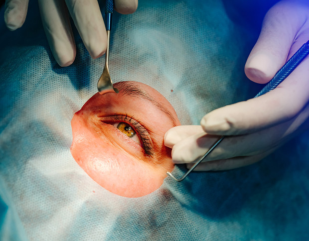
Indications for surgery are determined by more than one symptom. The doctor compares the patient's complaints, the rate of growth of the tumor, the position of the eye, visual function, and examination data.

In complex orbital cases, the decision is made by a team of specialists. It is important not only to remove the lesion but also to understand the scope of the intervention, the risks, and the subsequent follow-up after surgery.

When conservative treatment is no longer effective

Drug therapy, radiation therapy, or observation are not suitable for every patient. If the tumor is growing, destroying tissue, causing pain, or threatening vision, the doctor discusses surgical options. Orbital tumor removal is performed after confirmation of the indications.

Sometimes, surgical treatment of an orbital tumor becomes the primary method of disease control. This occurs when the tumor fails to respond to previously chosen therapy or requires removal based on morphological findings.

Tumors of the Orbit and Adjacent Tissues

Surgery may be necessary if the orbit, eyelids, lacrimal gland, conjunctiva, skin around the eye, or adjacent areas are affected. An orbital tumor can:

  • Displace the eye, limit movement.
  • Cause double vision, pain, or decreased vision.

If the diagnosis is confirmed, the doctor decides whether a limited orbital tumor removal is possible. If the tumor is deeply involved, the plan is expanded.

Surgical treatment of an orbital tumor depends on its structure. Benign, borderline, and malignant processes behave differently. In some situations, local excision is sufficient. In others, a more radical orbital surgery is necessary to avoid leaving an active lesion.

What symptoms require urgent consultation

Eyelid swelling is not always associated with an orbital tumor. However, there are signs that require an in-person examination. The orbit is located near the optic nerve and blood vessels. Therefore, significant changes require prompt diagnosis.

You should see a doctor immediately if you experience:

  • Rapid bulging of the eye
  • Sudden decrease in vision
  • Severe pain in the orbit
  • Double vision
  • Increasing swelling
  • Bleeding from the lesion

After consultation, the doctor will determine the cause of the symptoms and rule out conditions that may resemble a malignancy.

General information

How We Prepare a Patient for Surgery

Preparation begins with determining what exactly needs to be treated. Orbital surgery requires an understanding of the lesion's boundaries, proximity to the optic nerve, and the condition of the bony walls. It is important to determine whether the process has spread to adjacent tissues.

Examination: Ophthalmic Surgeon and Oncologist

During the consultation, the doctor assesses the eye's position, eyelids, conjunctiva, cornea, movement, and visual function. If a malignant or borderline process is suspected, an oncologist is involved in the analysis. The patient receives a unified examination plan and understands the purpose of each step.

Diagnostics: Orbital CT, Orbital MRI, biopsy, histology

Orbital CT helps assess the bony walls, dense inclusions, and the spread of the process to adjacent areas. Orbital MRI better shows:

  • Soft tissues
  • Muscles
  • Optic nerve
  • Relationship of the tumor to surrounding structures

If the nature of the tissue needs to be clarified, a biopsy is performed. Histology then reveals the cellular composition of the tumor and helps determine the treatment strategy.

Apparently similar changes can have different prognoses and different treatment plans. Orbital tumor removal is planned after comparing all data.

Planning the extent of the intervention and anesthesia

Before surgery, the doctor evaluates the patient's test results, chronic diseases, medications, and tolerance to medications. Anesthesia is selected based on the extent of the intervention, age, and general condition. The doctor discusses the intervention plan in person.

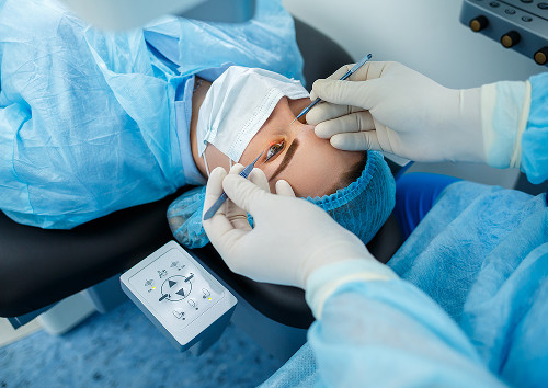
How We Prepare a Patient for Surgery

How the surgery is performed

In a modern clinical setting, orbital surgery is performed in the operating room, where the team controls access and the patient's condition. The surgeon works within a pre-calculated area. Certain decisions may be refined during the procedure if the actual lesion border differs from the imaging data.

Stages of surgery

Before surgery, the team verifies the surgical site, examination data, and the agreed-upon scope. The surgeon then accesses the affected tissue, isolates the lesion, removes the lesion, and evaluates the margins.

The specimen is sent for urgent or scheduled examination, as appropriate. After the main stage, the wound surface is formed, and a method for closure of the defect is selected.

In complex cases, a multidisciplinary approach is used. This is necessary if the lesion affects the skin, bone, paranasal sinuses, or deep orbital regions. The involvement of multiple specialists helps to plan treatment more accurately.

What is removed and what can be preserved

The extent of excision depends on the diagnosis, the boundaries of the lesion, and the involvement of adjacent tissues. Partial orbital exenteration may include removal of:

  • Damaged tissue
  • Part of the eyelid
  • Conjunctiva
  • Lacrimal gland
  • Other affected structures

If the eye and some function can be preserved without compromising safety, the doctor chooses a more gentle option. If the lesion has spread deeply, the plan changes.

Organ-preserving surgery requires a strict assessment of the boundaries. Tissue preservation should not leave the active lesion where it will continue to grow. The nuances of organ-preserving tactics are explained to the patient before the procedure.

How long does the surgery last and what is the follow-up process?

The duration depends on the extent of the lesion, the complexity of the approach, and the stage of the procedure. The exact time is determined after the examination, but it remains a guideline. After the surgery, the patient is observed in the ward and their general condition is monitored.

"In complex orbital cases, we always begin with an accurate diagnosis. Our goal is to choose the extent of the intervention that will ensure the safety of the treatment and, where possible, preserve healthy tissue," notes the ophthalmic surgeon.

How the surgery is performed

Recovery and Rehabilitation

The postoperative period depends on the extent of the removal and the reconstructive stage. Discomfort, soreness, changes in sensation, and dressing changes are possible in the first few days. The doctor assesses healing, explains care, and adjusts the prescription.

Early Postoperative Period

After surgery, the patient is initially monitored, especially after a major procedure. The team monitors the dressing, temperature, pain, and wound surface condition. If pain or weakness increases, notify the doctor.

Pain and Risk of Complications Management

Any orbital intervention involves tissue trauma. Possible complications:

  • Inflammation
  • Impaired healing
  • Changes in sensitivity
  • Cosmetic defect
  • Need for additional correction

Risks are mitigated by examination, careful technique, dressings, and regular monitoring. It is important for the patient to adhere to restrictions and attend checkups.

After the morphology results, the doctor will clarify the further plan. The morphology results may confirm complete removal or indicate the need for additional treatment. Sometimes, a consultation with a related specialist is necessary.

Orbital reconstruction, prosthetics, and follow-up

If the defect affects appearance or tissue protection, orbital reconstruction is discussed. It can be performed immediately or later, pending healing and analysis results. Rehabilitation includes dressings, scar monitoring, care training, and planning for the next steps. The patient receives a clear route, rather than disjointed recommendations.

Recovery and Rehabilitation

Why patients choose K+31

When orbital exenteration is needed in Moscow, the team's experience and diagnostic accuracy are crucial. The patient experiences the entire process from the initial consultation to post-operative follow-up within a single system.

Team approach and experience in ophthalmic oncology

Ophthalmic oncology treats tumors of the eye, orbit, and adjacent tissues. The structures responsible for vision, eye movement, and facial sensitivity are located nearby. This analysis reduces the risk of random decisions.

Accurate examination and individualized treatment strategy

Before surgery, we use examination, imaging data, tests, and morphological evaluation. Orbital tumors are assessed based on their location, structure, growth rate, and impact on visual function. Therefore, the same diagnosis may require different interventions in different patients.

For patients from other regions, Moscow is convenient because they can undergo diagnostics, surgery, and consultations with related specialists in a single city.

Patient support after surgery

Furthermore, after discharge, the patient remains under the supervision of specialists. The doctor monitors healing, morphological results, and recovery time. If additional correction is necessary, it is discussed after tissue stabilization. The second stop for patients is Moscow, where follow-up examinations and consultations can be combined.

Why patients choose K+31

Frequently Asked Questions

Patients often ask questions even before a consultation. Brief answers help clarify the treatment plan, but they don't replace an in-person examination.

Can the eye be saved?

Sometimes, saving the eye is possible if the lesion is limited and does not affect critical structures. Partial orbital exenteration is considered when there is a chance to remove the lesion without completely removing the orbital contents. However, there are no guarantees. The decision is made after imaging, examination, and tissue analysis.

How long does recovery take?

The recovery time depends on the extent of the removal, age, general condition, healing, and the need for plastic surgery. Recovery after a minor surgery is usually faster, while after a more extensive procedure, recovery takes longer. The doctor explains in advance which examinations are needed and when normal activities can be resumed. If additional treatment is planned, the schedule will be adjusted.

What are the possible risks and complications?

They depend on the extent of the surgery and the initial tissue condition. Pain, impaired healing, inflammatory reaction, changes in sensitivity, or cosmetic defects are possible. Postoperative monitoring helps to quickly respond to undesirable changes.

Is follow-up necessary?

Yes, follow-up is necessary. Even if the procedure went according to plan, the doctor needs to monitor the healing, morphological results, and the condition of the surrounding tissues. In the case of a tumor, follow-up examinations are important because the risk of recurrence is assessed dynamically. Ophthalmic oncology always requires discipline after treatment.
Conclusion

Conclusion

Partial orbital exenteration is a serious procedure used in situations where a dangerous lesion in the orbit must be removed while preserving healthy tissue, if medically justified. An orbital tumor cannot be assessed based on a single examination, as the lesion's boundaries are often determined only through a comprehensive diagnosis.

If you experience a mass in the orbit, bulging of the eye, pain, double vision, or decreased vision, it's best to seek a consultation without delay. K+31 specialists will assess your situation in person and explain which treatment option is best for you.

References

Our doctors

Karapetyan Arevik Samvelovna
Experience 12 years
Make an appointment
Karapetyan
Arevik Samvelovna
Ophthalmologist, Laser Surgeon
Batalina Larisa Vladimirovna
Experience 33 years
Make an appointment
Batalina
Larisa Vladimirovna
Deputy chief physician for ophthalmology, ophthalmic surgeon, leading specialist in laser vision correction
Amiryan Anush Gamletovna
Experience 28 years
Make an appointment
Amiryan
Anush Gamletovna
Ophthalmic oncologist surgeon
Medvedev Yulia Alexandrovna
Experience 11 years
Make an appointment
Medvedev
Yulia Alexandrovna
Ophthalmologist, Laser Surgeon
Rudkovskaya Elena Mikhailovna
Experience 38 years
Make an appointment
Rudkovskaya
Elena Mikhailovna
Ophthalmologist
Tegniryadnova Ekaterina Valerievna
Experience 20 years
Make an appointment
Tegniryadnova
Ekaterina Valerievna
Ophthalmologist, laser surgeon
Fadeeva Victoria Anatolievna
Experience 13 years
Make an appointment
Fadeeva
Victoria Anatolievna
Doctor ophthalmologist, laser surgeon
Shamsetdinova Leila Tagirovna
Experience 17 years
Make an appointment
Shamsetdinova
Leila Tagirovna
Pediatric ophthalmologist
Gavrilina Polina Dmitrievna
Experience 8 years
Make an appointment
Gavrilina
Polina Dmitrievna
Ophthalmologist, laser surgeon
Sugonyaeva Olga Yurievna
Experience 26 years
Make an appointment
Sugonyaeva
Olga Yurievna
Ophthalmologist, laser surgeon
Khlunovskaya Anna Nikolaevna
Experience 8 years
Make an appointment
Khlunovskaya
Anna Nikolaevna
Ophthalmologist
Akhiyarova Azalia Azatovna
Experience 4 years
Make an appointment
Akhiyarova
Azalia Azatovna
Ophthalmologist
Putintseva Polina Andreevna
Experience 6 years
Make an appointment
Putintseva
Polina Andreevna
Ophthalmologist
Agafonova Oksana Sergeevna
Experience 21 year
Make an appointment
Agafonova
Oksana Sergeevna
Ophthalmologist, pediatric
Kazennov Alexey Nikolaevich
Experience 19 years
Make an appointment
Kazennov
Alexey Nikolaevich
Ophthalmologist, ophthalmologist-surgeon
Petrova Maria Grigorievna
Experience 14 years
Make an appointment
Petrova
Maria Grigorievna
Ophthalmologist, ophthalmic surgeon
Mazmanyan Karen Akopovich
Experience 28 years
Make an appointment
Mazmanyan
Karen Akopovich
Ophthalmic surgeon
Suchkova Valeria Alekseevna
Experience 6 years
Make an appointment
Suchkova
Valeria Alekseevna
Ophthalmologist
Khubieva Salima Aslanbievna
Experience 7 years
Make an appointment
Khubieva
Salima Aslanbievna
Ophthalmologist, laser surgeon
Rytik Nina Petrovna
Experience 15 years
Make an appointment
Rytik
Nina Petrovna
Ophthalmologist
All specialists
2GIS Award
2GIS Award

This award is given to clinics with the highest ratings according to user ratings, a large number of requests from this site, and in the absence of critical violations.

«Good place» according to Yandex

This award is given to clinics with the highest ratings according to user ratings. It means that the place is known, loved, and definitely worth visiting.

Our doctors are laureates of the ProDoctors Award

The ProDoctors portal collected 500 thousand reviews, compiled a rating of doctors based on them and awarded the best. We are proud that our doctors are among those awarded.

Make an appointment at a convenient time on the nearest date

Price

Reception
Price
Initial Ophthalmologist Appointment
from 6 450 ₽
Corneal Collagen Crosslinking with Ultraviolet Irradiation
from 52 500 ₽
Femtosecond Laser-Assisted Intrastromal Segment Implantation
from 200 000 ₽

Other services

Laser vision correction Phacoemulsification of cataract with IOL implantation of any complexity Vitreoretinal surgeries of any complexity (vitrectomy) Laser photocoagulation of the retina during tears and degenerative changes in the retina Laser photocoagulation for diabetic retinopathy Intravitreal administration of Anti-VEGF drugs, hormonal implants Antiglaucomatous operations using various drains YAG laser dissection of the posterior capsule for secondary cataract Cataract treatment Treatment of glaucoma Retinal dystrophic changes Vitreous pathology Injury to the eye Post-traumatic and postoperative complications Myopia treatment Farsightedness Treatment of astigmatism Retinal disinsertion Non-contact laser procedure SMARTSURFACE Keratoplasty Femto SUPER LASIK Laser coagulation of the retina of the eye treatment on Ellex Integre Pro Scan Treatment of the retina with the Navilas laser system Femtolaser Cataract Removal Selection of children's night lenses Laser vision correction Clear Офтальмолог-онколог в Москве Treatment of keratoconus Laser coagulation of the retina Refractometry Retrobulbar injections Restoration of vision Lacrimal ducts Eye wash Synoptophore Forbis ESOM Pediatric ophthalmologist Ophthalmologist Glasses Biomicroscopy Night lenses Photo of an eye Eyelid suturing Selection of lenses Retinal laser Vision test Vitrectomy Vitrectomy Vitrectomy Eye pressure Ophthalmoscopy Fundus Gonioscopy Discision Orbitotomy Ophthalmodiagnostics OCT of the optic disc Keratoplasty Crystalline lens replacement Lacrimal duct Eye biometrics Angle of strabismus Lacrimal point IOL implantation Heterophoria Refraction Needling pillows Sclerectomy Micropulse laser Eyelid massage Intravitreal injections Iridoplasty Accommodation Bacterial culture Ophthalmology Cyclophotocoagulation Laser trabeculoplasty Laser stimulation Makdel Iridotomy Goniopuncture Corneal crosslinking Perimetry Pachymetry Angio-OCT Tubular test Color vision Convergence test Autofluorescence Eye mobility Corneal diagnostics Corneal transplant Skiascopy Strabismus Injection in the eye Tonometry Schirmer's test Trabeculotomy Diaphanoscopy Foreign body Foreign body Eyelid neoplasms Retinal tear Pterygium Chalazion Pneumatic retinopexy Glaucoma Ultrasound of the eye Eye biometrics Improving vision Cataract removal An attack of glaucoma Night lenses Eyelid ptosis Vision correction Angiography of the eye Retinoblastoma Laser coagulation PRK Evisceration of the eye Exophthalmometry PresbyLasik Removal of the lens Retinal thrombosis Nystagmus Lacrimal duct Eye removal Optic nerve Eyelash removal Neoplasms of the eyelids Myopia Autorefractometry Anti-VEGF Iris melanoma Glaucoma treatment AMVO-01
Make an appointment

Appointment to the doctor

Fill out the form, our managers will contact you within 15 minutes

Reviews

The reception was professional and unobtrusive. Additional examinations and diagnostics were carried out using modern equipment. During the consultation and examination, Polina Dmitrievna explained everything in detail and intelligibly, gave me clear recommendations, and answered all questions in detail. Polina Dmitrievna is a polite, attentive and simply amazing doctor. The clinic as a whole enjoys the atmosphere, responsiveness of the staff and the professionalism of the specialists.
11.07.2026
Nikolay K.
Rudkovskaya E.M. is an excellent doctor, if you need an ophthalmologist, I go to her. A very competent specialist. Explains everything by diagnosis. Comprehensively evaluates the condition, checking all the parameters.
11.07.2026
Vera S.
Wonderful doctor!
10.07.2026
Natalya P.
An excellent specialist. I am very pleased with the reception. Thanks to the doctor
08.07.2026
Svetlana U.
I would like to express my sincere gratitude to all the staff of the K+31 clinic. Special thanks to the doctors of the ophthalmology department, Ekaterina V. Tegniryadnova, who has been seeing me for a long time, and Maria Grigorievna Petrova, who operated on me for a lens replacement. Everything went great! I would also like to mention the work of Diana's ophthalmology manager. I received comprehensive and timely answers to all my questions. THANK you so much to all of you!
I am very pleased with the reception, smart doctor, thank you By +31.
26.06.2026
Irina T.
The Miracle Doctor
24.06.2026
Anush D.
Почему К+31?
К + 31 — full-cycle multidisciplinary medical centers, including the possibility of providing medical services of European quality level.
К + 31 — are leading doctors and diagnostics using high-tech equipment from world manufacturers (Karl Storz, Olympus, Siemens, Toshiba, Bausch&Lomb, Technolas, Zeiss, Topcon).
К + 31 — is ethical. The staff of K+31 clinics maintain open relationships with patients and partners. An individual approach to each patient is the basis of our service standards.
К + 31 — is modernity. On call 24/7: call center operators will answer your questions at any time and book you an appointment with doctors. Contact us by phone, through the feedback form on the website and Max.

Our clinics

Address K+31 Lobachevskogo 2

st. Lobachevskogo, 42/7

Contacts

+7 499 999-31-31

Opening hours

Mon-Fri: 08:00 – 21:00
Weekend: 09:00 – 19:00

Address K+31 on Lobachevskogo

st. Lobachevskogo, 42/4

Contacts

+7 499 999-31-31

Opening hours

Mon-Fri: 08:00 – 21:00
Weekend: 09:00 – 19:00

Address K+31 Petrovskie Vorota

1st Kolobovsky pereulok, 4

Contacts

+7 499 999-31-31

Opening hours

Mon-Fri: 08:00 – 21:00
Sat-Sun: 09:00 – 19:00

Address K+31 West

Orshanskaya, 16/2; Ak. Pavlova, 22

Contacts

+7 499 999-31-31

Opening hours

Mon-Fri: 08:00 – 21:00
Sat-Sun: 09:00 – 18:00
K+31 on Lobachevskogo
Didn't find the service you were looking for?

Экстренная помощь