Corneal Examination: Diagnostic Methods, Indications, and Examination Results

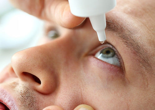
The cornea is the first to react to dryness, infection, microtrauma, and contact lenses. Its shape, thickness, and transparency determine the clarity of vision and the safety of treatment.

At K+31, corneal examinations are comprehensive: the doctor compares the patient's complaints, instrument data, and the condition of the eye.

Best specialists
Best
specialists
Expert equipment
Expert
equipment
Advanced diagnostic treatment
Advanced diagnostic
treatment
Download the application “Personal Account K+31”
Everything about your health in one click!
Subscribe to social networks K+31
To keep up to date with current offers and receive useful advice about your health.

What is a corneal examination and why is it necessary?

The cornea is the transparent front layer of the eye through which light passes into the eye. Changes in its surface can cause blurriness, glare, a gritty sensation, or unstable vision.

A corneal examination helps assess the shape, thickness, transparency, and internal layers of the tissue. The doctor looks for inflammation, scarring, swelling, thinning, and the effects of injury or surgery.

What we evaluate during the examination:

  • Corneal transparency
  • Corneal thickness
  • Corneal curvature
  • Tear film condition
  • Corneal endothelium
  • Scars, opacities, thinning, or inflammation

Based on this information, the doctor chooses the next step.

What is a corneal examination and why is it necessary?

When do we recommend a corneal examination?

Corneal examinations are prescribed for complaints, before surgeries, and after injuries, burns, inflammatory diseases, and interventions. If vision suddenly worsens, it's best not to wait for the scheduled appointment.

Examination before surgery and correction selection

Before surgery, it's important to know whether the tissue is stable and thick enough. If laser vision correction, cataract removal, or a prescription for complex lenses is planned, the doctor may prescribe:

  • Keratometry
  • Keratotopography
  • Corneal pachymetry
  • Tomographic scanning

This allows the doctor to assess risks in advance and make a decision after an in-person examination.

Complaints that require an appointment for diagnostics

An ophthalmologist should be consulted if the patient experiences pain, redness, photophobia, tearing, stinging, glare, a foreign body sensation, or decreased visual acuity. The doctor will ask whether the patient wears contact lenses and whether they have had any infections, injuries, surgeries, or chemical irritation.

The cornea is sensitive to dryness, inflammation, and improper lens fit. Without an examination, it's difficult to determine whether the cause is on the surface, deep within the tissue, or elsewhere in the visual system.

Monitoring after Treatment and Injuries

After inflammation, a burn, a foreign body, surgery, or prolonged irritation, it's important to understand how healing is progressing. The doctor evaluates the corneal surface, swelling, sensitivity, and cloudiness, if any, that developed after the injury.

Repeat corneal examinations show progress and help adjust monitoring schedules.

What methods of corneal examination do we use?

The method is chosen by the ophthalmologist after a conversation and initial examination. Below are the main methods that help assess the anterior segment of the eye.

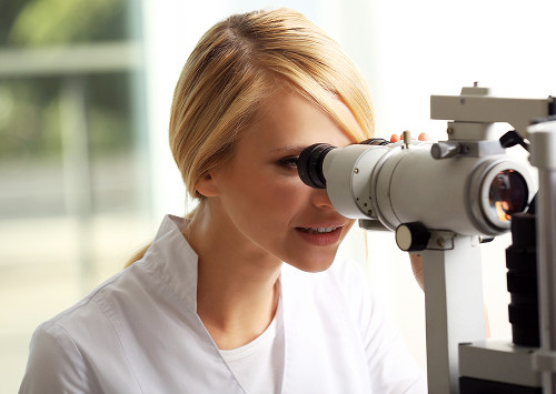
Slit Lamp Examination

A slit lamp provides a magnified image of the anterior structures of the eye. During the examination, biomicroscopy is performed: the doctor can see the surface, tear film, epithelium, stromal layer, and any opacities.

A slit lamp also helps detect microdefects after staining and determine whether additional measurements are needed.

Keratometry

Keratometry measures the curvature of the central zone of the anterior surface. This method is important for astigmatism, preparation for surgery, and selection of optical correction.

If the results are unstable, an extended surface map is prescribed.

Corneal Pachymetry

Corneal pachymetry measures tissue thickness at selected points or maps its distribution. This measurement is important when suspecting keratoconus, before refractive surgery, for edema, and for post-treatment monitoring.

A thin zone, asymmetry, or dynamic changes in the data help determine whether more frequent monitoring is necessary.

Keratotopography and Keratotomography

Keratotopography creates a map of the anterior surface and shows the distribution of refractive power. Corneal topography is useful for irregular astigmatism, poor lens tolerance, and suspected shape changes.

Corneal tomography evaluates the anterior and posterior surfaces, thickness distribution, and signs of thinning. These data help identify early keratoconus when a routine eye exam fails to explain the symptoms.

Anterior Segment Optical Coherence Tomography

Anterior segment optical coherence tomography creates a layered image of tissue without incisions or eye contact. This method helps assess the depth of opacities, scars, swelling, and post-operative conditions.

OCT is important when the physician needs to understand not only the presence of a change but also its location.

Endothelial Microscopy

Endothelial microscopy evaluates the cells of the inner layer, which maintains normal tissue water balance. If the cells are few or altered, the risk of postoperative edema increases.

The corneal endothelium is especially important before surgery, after keratoplasty, and in cases of dystrophies and chronic edema.

Confocal Microscopy

Confocal microscopy provides a layered image of tissue at the cellular level. This method is used according to the following indications:

  1. Complex inflammation
  2. Suspected specific lesion
  3. Dystrophies or unclear pain

The decision to prescribe is made by the physician after assessing the patient's complaints and previous data.

Before choosing a method, the physician compares several parameters. For the patient, differences are easier to see through comparison.

Method What it shows When it's needed Features
Biomicroscopy surface, transparency, inflammation initial examination quickly
Corneal pachymetry tissue thickness keratoconus, preparation for surgery indicator Thickness
Keratometry Central curvature Astigmatism, optical calculation Evaluates refraction
Corneal topography Shape and distribution of curvature Lens fitting Non-contact
Endothelial microscopy Inner layer cells Surgeries, dystrophies Tissue reserve

This table does not replace a consultation. The doctor selects a set of tests after the examination.

General information

When a corneal examination cannot be postponed

A scheduled appointment is not always appropriate. If symptoms are severe, consult a doctor immediately.

Pain, redness, and photophobia

Severe pain, redness, photophobia, and excessive tearing may indicate corneal inflammation, erosion, keratitis, trauma, or lens complications. Waiting several days is dangerous.

Decreased vision and a "foggy" sensation

Sudden decreased vision, haze, glare, a filmy sensation, or severe swelling require an in-person examination. The doctor determines whether the cornea's transparency has changed, whether there is an infiltrate or scar.

Trauma, burn, or severe eye irritation

After exposure to a chemical, wood shavings, twig, sharp object, or hot steam, the eye should be examined by a specialist. The doctor assesses the depth of the defect, the condition of the eyelids, and the risk of infection.

When a corneal examination cannot be postponed

How is the examination performed?

A typical appointment begins with complaints, anamnesis, and a vision test. The doctor then conducts an examination and prescribes the necessary measurements. At K+31, patients receive results and recommendations in one visit unless urgent additional care is needed.

Is preparation necessary?

Special preparation is not always necessary. If you wear contact lenses, use eye drops, or have recently had an infection or surgery, please let us know in advance.

How long does the examination take?

The duration depends on the scope of the diagnostic work. The initial examination is quicker, while the advanced program with surface mapping, OCT, and endothelial assessment takes longer. After dilating drops, temporary blurred vision and light sensitivity are possible.

What does the patient feel during the procedure?

Most methods are painless. For contactless measurements, you should calmly look at the mark.

How is the examination performed?

What does the diagnostic show and how do we interpret the results?

The results are reviewed together: complaints, visual acuity, surface condition, charts, thickness, microscopy data, and the patient's history. Therefore, it is better to conduct a comprehensive corneal examination rather than collecting individual numbers without a doctor's interpretation.

Corneal Thickness and Curvature

Corneal curvature affects light refraction and image quality. Its change can be associated with astigmatism, lenses, surgery, or tissue disease. If localized thinning is present, the doctor assesses the risk of progression.

Tissue Transparency and Structure

Corneal clouding can be superficial or deep. The treatment strategy depends on the depth:

  1. Observation
  2. Treatment of inflammation
  3. Correction of dryness
  4. Additional tests
  5. Surgical treatment option

Optical coherence tomography helps to clarify the location of the lesion.

Endothelial status and signs of pathology

The inner layer maintains tissue transparency by controlling fluid. If cells are damaged, the risk of edema increases. Based on microscopy data, the ophthalmologist explains whether monitoring, restrictions, or preparation are needed before surgery.

What does the diagnostic show and how do we interpret the results?

Why patients choose K+31 for corneal examinations

For patients, it's not just the equipment that matters, but also the doctor's interpretation. At K+31, diagnostics are structured so that people understand what was found, what it means, and what the safest course of action is.

Comprehensive diagnostics in one visit

We can perform several examinations during a single visit if needed. This format helps us quickly compare the shape, thickness, transparency, and condition of the cornea's inner layers.

Ophthalmologists with practical experience

Our ophthalmologists treat complaints of varying complexity: from dryness and contact lens problems to the consequences of surgeries, injuries, and degenerative diseases. The doctor explains the results in medical terms, but without unnecessary pressure.

Modern equipment and accurate interpretation

The devices produce maps, images, and numerical values. The doctor assigns clinical significance to this data: they identify the norm, an individual characteristic, or a sign of a disease. Therefore, corneal diagnostics at our clinic always involve an in-person assessment.

Clear recommendations after the examination

After the appointment, the patient receives an explanation of the results and a further plan. Sometimes observation is sufficient, while other times treatment, lens correction, preparation for surgery, or a consultation with a related specialist are necessary.

"In our practice, corneal examination often helps us identify changes that are not noticeable during a routine vision exam. We use several methods in combination to develop a precise action plan," says the ophthalmologist.

Why patients choose K+31 for corneal examinations

Frequently Asked Questions

What does the examination reveal?

The examination helps assess the shape, thickness, curvature, transparency, and overall condition of the tissue. The doctor can also detect inflammation, swelling, scarring, dystrophies, and thinning.

Is a corneal examination painful?

Usually not. Most procedures are quick, with minimal or no contact with the eye.

Is any preparation necessary for a corneal examination?

Sometimes, preparation is necessary. You may need to temporarily remove your lenses before certain measurements, but it's best to do so as directed by your doctor. Please inform your doctor about any eye drops, injuries, or surgeries.

When is a corneal examination prescribed?

A corneal examination is prescribed for decreased vision, pain, redness, suspected thinning, after injuries, before surgery, when fitting lenses, and for post-treatment monitoring. Urgent consultation is necessary for burns, severe pain, or sudden vision loss.

Conclusion: When it is important not to delay a visit

Conclusion: When it is important not to delay a visit

A corneal examination is recommended if your vision has become unstable, you experience pain, photophobia, redness, blurred vision, or discomfort from your lenses. This also applies to preparation for surgery, post-injury monitoring, and any changes following inflammation.

At "K+31," a doctor conducts an examination, selects appropriate methods, and explains the results in understandable language. Schedule a consultation if symptoms are interfering with your vision or have appeared suddenly.

Our doctors

Karapetyan Arevik Samvelovna
Experience 12 years
Make an appointment
Karapetyan
Arevik Samvelovna
Ophthalmologist, Laser Surgeon
Batalina Larisa Vladimirovna
Experience 33 years
Make an appointment
Batalina
Larisa Vladimirovna
Deputy chief physician for ophthalmology, ophthalmic surgeon, leading specialist in laser vision correction
Amiryan Anush Gamletovna
Experience 28 years
Make an appointment
Amiryan
Anush Gamletovna
Ophthalmic oncologist surgeon
Medvedev Yulia Alexandrovna
Experience 11 years
Make an appointment
Medvedev
Yulia Alexandrovna
Ophthalmologist, Laser Surgeon
Rudkovskaya Elena Mikhailovna
Experience 38 years
Make an appointment
Rudkovskaya
Elena Mikhailovna
Ophthalmologist
Tegniryadnova Ekaterina Valerievna
Experience 20 years
Make an appointment
Tegniryadnova
Ekaterina Valerievna
Ophthalmologist, laser surgeon
Fadeeva Victoria Anatolievna
Experience 13 years
Make an appointment
Fadeeva
Victoria Anatolievna
Doctor ophthalmologist, laser surgeon
Shamsetdinova Leila Tagirovna
Experience 17 years
Make an appointment
Shamsetdinova
Leila Tagirovna
Pediatric ophthalmologist
Gavrilina Polina Dmitrievna
Experience 8 years
Make an appointment
Gavrilina
Polina Dmitrievna
Ophthalmologist, laser surgeon
Kovaleva Olesya Alexandrovna
Experience 7 years
Make an appointment
Kovaleva
Olesya Alexandrovna
Ophthalmologist, laser surgeon
Sugonyaeva Olga Yurievna
Experience 26 years
Make an appointment
Sugonyaeva
Olga Yurievna
Ophthalmologist, laser surgeon
Khlunovskaya Anna Nikolaevna
Experience 8 years
Make an appointment
Khlunovskaya
Anna Nikolaevna
Ophthalmologist
Akhiyarova Azalia Azatovna
Experience 4 years
Make an appointment
Akhiyarova
Azalia Azatovna
Ophthalmologist
Putintseva Polina Andreevna
Experience 6 years
Make an appointment
Putintseva
Polina Andreevna
Ophthalmologist
Agafonova Oksana Sergeevna
Experience 21 year
Make an appointment
Agafonova
Oksana Sergeevna
Ophthalmologist, pediatric
Kazennov Alexey Nikolaevich
Experience 19 years
Make an appointment
Kazennov
Alexey Nikolaevich
Ophthalmologist, ophthalmologist-surgeon
Petrova Maria Grigorievna
Experience 14 years
Make an appointment
Petrova
Maria Grigorievna
Ophthalmologist, ophthalmic surgeon
Mazmanyan Karen Akopovich
Experience 28 years
Make an appointment
Mazmanyan
Karen Akopovich
Ophthalmic surgeon
Suchkova Valeria Alekseevna
Experience 6 years
Make an appointment
Suchkova
Valeria Alekseevna
Ophthalmologist
Khubieva Salima Aslanbievna
Experience 7 years
Make an appointment
Khubieva
Salima Aslanbievna
Ophthalmologist, laser surgeon
Rytik Nina Petrovna
Experience 15 years
Make an appointment
Rytik
Nina Petrovna
Ophthalmologist
All specialists
2GIS Award
2GIS Award

This award is given to clinics with the highest ratings according to user ratings, a large number of requests from this site, and in the absence of critical violations.

«Good place» according to Yandex

This award is given to clinics with the highest ratings according to user ratings. It means that the place is known, loved, and definitely worth visiting.

Our doctors are laureates of the ProDoctors Award

The ProDoctors portal collected 500 thousand reviews, compiled a rating of doctors based on them and awarded the best. We are proud that our doctors are among those awarded.

Make an appointment at a convenient time on the nearest date

Price

Reception
Price
Initial Ophthalmologist Appointment
from 6 450 ₽
Corneal Collagen Crosslinking with Ultraviolet Irradiation
from 52 500 ₽
Femtosecond Laser-Assisted Intrastromal Segment Implantation
from 200 000 ₽

Other services

Laser vision correction Phacoemulsification of cataract with IOL implantation of any complexity Vitreoretinal surgeries of any complexity (vitrectomy) Laser photocoagulation of the retina during tears and degenerative changes in the retina Laser photocoagulation for diabetic retinopathy Intravitreal administration of Anti-VEGF drugs, hormonal implants Antiglaucomatous operations using various drains YAG laser dissection of the posterior capsule for secondary cataract Cataract treatment Treatment of glaucoma Retinal dystrophic changes Vitreous pathology Injury to the eye Post-traumatic and postoperative complications Myopia treatment Farsightedness Treatment of astigmatism Retinal disinsertion Non-contact laser procedure SMARTSURFACE Keratoplasty Femto SUPER LASIK Laser coagulation of the retina of the eye treatment on Ellex Integre Pro Scan Treatment of the retina with the Navilas laser system Femtolaser Cataract Removal Selection of children's night lenses Laser vision correction Clear Офтальмолог-онколог в Москве Treatment of keratoconus Laser coagulation of the retina Refractometry Retrobulbar injections Restoration of vision Lacrimal ducts Eye wash Synoptophore Forbis ESOM Pediatric ophthalmologist Ophthalmologist Glasses Biomicroscopy Night lenses Photo of an eye Eyelid suturing Selection of lenses Retinal laser Vision test Vitrectomy Vitrectomy Vitrectomy Eye pressure Ophthalmoscopy Fundus Gonioscopy Discision Orbitotomy Ophthalmodiagnostics OCT of the optic disc Keratoplasty Crystalline lens replacement Lacrimal duct Eye biometrics Angle of strabismus Lacrimal point IOL implantation Heterophoria Refraction Needling pillows Sclerectomy Micropulse laser Eyelid massage Intravitreal injections Iridoplasty Accommodation Bacterial culture Ophthalmology Cyclophotocoagulation Laser trabeculoplasty Laser stimulation Makdel Iridotomy Goniopuncture Corneal crosslinking Perimetry Pachymetry Angio-OCT Tubular test Color vision Convergence test Autofluorescence Eye mobility Corneal transplant Skiascopy Injection in the eye Tonometry Schirmer's test Trabeculotomy Diaphanoscopy Foreign body
Make an appointment

Appointment to the doctor

Fill out the form, our managers will contact you within 15 minutes

Reviews

Super doctor
29.06.2026
Aleksey K.
I would like to express my sincere gratitude to all the staff of the K+31 clinic. Special thanks to the doctors of the ophthalmology department, Ekaterina V. Tegniryadnova, who has been seeing me for a long time, and Maria Grigorievna Petrova, who operated on me for a lens replacement. Everything went great! I would also like to mention the work of Diana's ophthalmology manager. I received comprehensive and timely answers to all my questions. THANK you so much to all of you!
I am very pleased with the reception, smart doctor, thank you By +31.
26.06.2026
Irina T.
The Miracle Doctor
24.06.2026
Anush D.
I underwent preoperative preparation before laser correction at Azaliya Azatovna, I was satisfied! A very sensitive and understanding doctor. I drew schematically how the operation would take place, it was interesting and the fear went away. She also performed postoperative examinations. I wish you grateful patients and success in carrying out laser corrections. I will recommend it to my friends and family.
23.06.2026
Ekaterina Sh.
A very qualified doctor! Attentive and caring!
20.06.2026
Olga Ch.
Olesya Alexandrovna is an excellent specialist, listened attentively and carried out the necessary diagnostics, answered all questions in detail and kindly and gave recommendations. I plan to see Dr. Kovaleva in the future.
18.06.2026
Anton Sh.
Почему К+31?
К + 31 — full-cycle multidisciplinary medical centers, including the possibility of providing medical services of European quality level.
К + 31 — are leading doctors and diagnostics using high-tech equipment from world manufacturers (Karl Storz, Olympus, Siemens, Toshiba, Bausch&Lomb, Technolas, Zeiss, Topcon).
К + 31 — is ethical. The staff of K+31 clinics maintain open relationships with patients and partners. An individual approach to each patient is the basis of our service standards.
К + 31 — is modernity. On call 24/7: call center operators will answer your questions at any time and book you an appointment with doctors. Contact us by phone, through the feedback form on the website and Max.

Our clinics

Address K+31 Lobachevskogo 2

st. Lobachevskogo, 42/7

Contacts

+7 499 999-31-31

Opening hours

Mon-Fri: 08:00 – 21:00
Weekend: 09:00 – 19:00

Address K+31 on Lobachevskogo

st. Lobachevskogo, 42/4

Contacts

+7 499 999-31-31

Opening hours

Mon-Fri: 08:00 – 21:00
Weekend: 09:00 – 19:00

Address K+31 Petrovskie Vorota

1st Kolobovsky pereulok, 4

Contacts

+7 499 999-31-31

Opening hours

Mon-Fri: 08:00 – 21:00
Sat-Sun: 09:00 – 19:00

Address K+31 West

Orshanskaya, 16/2; Ak. Pavlova, 22

Contacts

+7 499 999-31-31

Opening hours

Mon-Fri: 08:00 – 21:00
Sat-Sun: 09:00 – 18:00
K+31 on Lobachevskogo
Didn't find the service you were looking for?

Экстренная помощь