Angio-OCT (OCT angiography) of the eye in Moscow
OCT Angiography is a modern diagnostic method that helps assess the retinal vascular network without injections or contrast. This examination allows the doctor to detect changes in microcirculation, evaluate the macula, capillaries, and individual structures of the optic nerve even before the onset of significant symptoms.
At K+31, we use modern equipment for precise vascular visualization and thoroughly explain the results to the patient immediately after diagnosis.

specialists

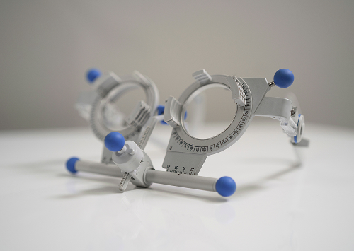
equipment

treatment


When do we recommend Angio-OCT?

This examination is prescribed not only for existing diagnoses but also for complaints that may be related to retinal blood flow problems.
Complaints that warrant an examination
Indications for the procedure:
- Decreased visual acuity
- Distorted lines or a "spot" in the center of vision
- Diabetes
- Suspected glaucoma
- Disease monitoring
- Post-treatment monitoring
Flashes of light, decreased central vision, and difficulty reading may also be reasons for an examination.

Diseases and conditions that this method helps monitor
Octanoscopic angiography of the eye is actively used to monitor patients with chronic diseases and vascular disorders.
This method helps monitor:
- Diabetic retinopathy
- Age-related macular degeneration
- Glaucoma
- Vascular pathologies
- Macular edema
- Optic nerve pathology
In some cases, this method can detect early signs of problems even before significant vision loss occurs.
How the study is conducted
For most patients, the procedure is relaxing and doesn't cause significant discomfort. We explain each diagnostic step in advance and answer any questions before the examination.
Is preparation necessary?
No preparation is necessary. Sometimes, your ophthalmologist may recommend dilating your pupils with drops if needed for a more accurate scan.
It's important to inform your doctor about any eye conditions, treatments, and previous examinations.
How the procedure is performed
The patient is positioned in front of the machine and fixes their gaze on a light target. A series of images and a tomography of the ocular structures are then taken.
The scan takes little time and is performed painlessly and without contrast. This is why many patients choose angio-OCT of the eye in Moscow as a more comfortable examination option compared to invasive methods.
The quality of the images depends on the transparency of the ocular media and the patient's ability to fixate their gaze during the examination.
What does the patient receive after diagnosis?
After the examination, the doctor interprets the images and explains the results in simple terms. The patient receives diagnostic data, recommendations, and an algorithm for monitoring or treatment.
"We recommend Angio-OCT when it's important to quickly and without contrast evaluate the retinal vessels and identify early abnormalities. In our practice, this is especially valuable: the patient receives a clear explanation of the results, and we receive a precise basis for further action."
General information
Advantages of Angio-OCT in our clinic
No contrast and painless
One of the main advantages of this method is that the examination is performed without injections. Contrast is also not required.
This approach makes the examination more comfortable and helps with diagnostics even when regular retinal monitoring is necessary.
Highly detailed image
Modern equipment allows us to obtain high-quality images and assess the presence of pathologies at the earliest stages.
The doctor can see the condition of the capillaries, microcirculation characteristics, and changes in the macula. This is especially important in the early diagnosis of vascular diseases of the eye.
Fast results and expert interpretation
Experienced doctors work at the K+31 clinic. After the diagnosis, the ophthalmologist immediately explains the examination results and answers the patient's questions.
We focus not only on diagnostic accuracy but also on clear interpretation of the images.


Comparison of Angio-OCT with other diagnostic methods
| Method | What it shows | Is contrast needed? | How long it lasts | When it is used |
|---|---|---|---|---|
| Angio-OCT of the eye | Retinal structure and vessels | No | ~5–10 min | Diagnosis of vascular changes, maculopathy, glaucoma |
| Classical OCT | Structure of retinal layers | No | ~5 min | Detection of edema, tears, and AMD |
| Fluorescence angiography | Vascular patency and ischemic zones | Yes | ~20–30 min | Severe diabetic retinopathy, thrombosis |
Angio-OCT and conventional OCT
The main difference is the ability to evaluate not only tissue structure but also the vascular network. Therefore, the methods often complement each other.
Angio-OCT and fluorescein angiography
Fluorescein angiography remains an important diagnostic method in complex cases, but requires the administration of contrast.
Angio-OCT provides vascular data without injections.
When additional tests are needed
Sometimes, an ophthalmologist may recommend additional diagnostic methods to clarify the diagnosis or evaluate complex changes.
Why patients choose us

Experienced ophthalmologists
At our clinic, diagnostics are performed by specialists with experience in vascular assessment.
Each ophthalmologist considers not only the images but also the overall clinical picture.
Modern equipment
We use modern equipment for comprehensive diagnostics and precise visualization of the ocular vascular network.
Convenient appointment and patient follow-up
Patients receive results quickly and can immediately discuss treatment issues with the doctor. This helps avoid delays in examinations and ensure timely vision monitoring.
FAQ
What does an Angio-OCT scan of the eye reveal?
Is this procedure painful?
How does Angio-OCT differ from conventional OCT and fluorescein angiography?

Our doctors


This award is given to clinics with the highest ratings according to user ratings, a large number of requests from this site, and in the absence of critical violations.

This award is given to clinics with the highest ratings according to user ratings. It means that the place is known, loved, and definitely worth visiting.

The ProDoctors portal collected 500 thousand reviews, compiled a rating of doctors based on them and awarded the best. We are proud that our doctors are among those awarded.
Make an appointment at a convenient time on the nearest date
Price
Other services

Appointment to the doctor
Reviews


Our clinics

Application “Personal Account K+31”


What is Angio-OCT and how does it differ from conventional OCT?
OCT angiography of the eye is a tomography method that allows the doctor to obtain images of the retinal vasculature and assess blood flow in small vessels without injecting dye. This noninvasive examination is widely used in ophthalmology for the early detection of vascular pathologies.
This method reveals the tissue structure and layer thickness and helps analyze microcirculation, capillary function, and the blood supply to the macula.
What retinal layers and vessels are visible in this examination?
During scanning, the doctor evaluates several levels of the vascular network. The images show the superficial and deep vascular plexuses, choriocapillaries, the macula, and specific areas of the optic nerve.
This retinal examination helps identify pathological changes that are not always visible during a standard examination, such as areas of ischemia, early neovascularization, or microcirculatory disorders.
This is especially important if diabetes, vascular disorders, and age-related macular degeneration are suspected.
Why is Angio-OCT considered a non-invasive diagnostic?
The main difference of this method is the absence of intravenous dye injection. This is why Angio-OCT is considered a comfortable and gentle diagnostic option.
The patient does not require a contrast injection, and the procedure itself takes little time. This is especially important for people who have difficulty tolerating invasive examinations.
It is important to understand that even modern non-invasive early diagnostics does not replace a consultation with an ophthalmologist and a comprehensive examination.