
Closed-angle glaucoma: symptoms, diagnosis and treatment
Closed-angle glaucoma develops when fluid drainage within the eye is blocked. Pressure can rise quickly, causing pain and a sharp deterioration in vision.
At K+31, we assess your condition immediately: we conduct an examination, explain the results, and develop a treatment plan.

specialists

equipment

treatment






Why does angle-closure glaucoma develop?
The disease is associated with structural features of the eye and factors that increase angle closure. The risk increases:
- With a small anterior chamber
- Age-related changes
- A specific position of the iris and lens
Sometimes the problem is discovered during a routine eye exam, when there are no significant complaints.

-
Anatomically narrow anterior chamber angle
This means the space for fluid drainage is limited. When the pupil dilates, the iris can block the drainage area, causing intraocular pressure to rise. The patient may experience pain, fullness, or clouding.
-
Factors that can trigger an attack
With narrow-angle glaucoma, an attack sometimes begins after pupil dilation. This can occur in the dark, during intense emotional stress, or after taking certain medications or diagnostic drops. If narrow-angle glaucoma has been suspected, it's best to discuss the list of regular medications with a specialist.
Symptoms of angle-closure glaucoma
Symptoms depend on the rate of angle closure. In the acute form, complaints appear suddenly. In the chronic form, symptoms are milder, so the patient may attribute them to fatigue or age for a long time.
Acute attack: how it manifests
Most often, an attack begins with severe discomfort in one eye. Possible symptoms include:
- Eye pain
- Redness of the eye
- Foggy vision
- Rainbow halos around lights
Nausea and vomiting sometimes occur. During an attack, intraocular pressure may be elevated.
Due to headache and nausea, an attack is sometimes mistaken for another condition. An urgent examination and blood pressure measurement are necessary.
Chronic course: hidden signs
Complaints may be mild. The patient notices periodic blurring, a feeling of pressure, decreased clarity in the evening, and discomfort in low light. During this time, visual field may gradually suffer.
When visual field narrowing occurs, some of the changes may already be permanent. Therefore, recurring episodes of blurring are best assessed in person.
When you need urgent help
There are signs that prevent you from waiting for a scheduled consultation. They may indicate a sharp increase in pressure and a threat to the visual structures. They should be especially carefully monitored if a narrow angle has been detected.
Symptoms that prevent you from waiting:
- Sharp eye pain
- Sudden blurred vision
- Rainbow halos around light
- Redness of the eye
- Nausea or headache
- Rapid deterioration of vision
If these symptoms appear, emergency care is needed. Before the examination, do not try to prescribe eye drops, warm the eye, or wait for the pain to subside. Without a diagnosis, it is impossible to safely distinguish an attack from other conditions.
How do we diagnose angle-closure glaucoma?
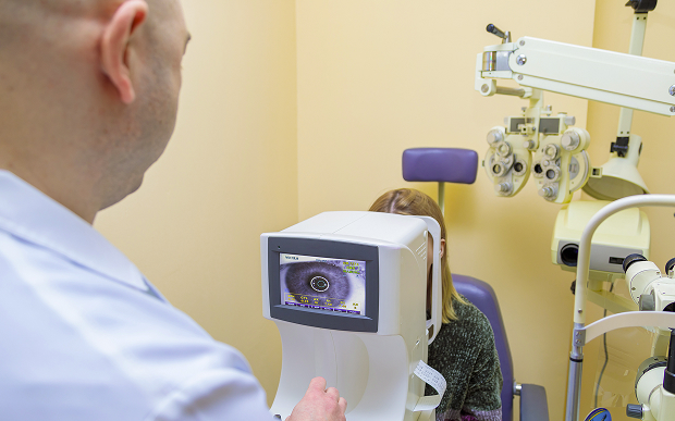
The diagnosis of angle-closure glaucoma begins with complaints and an initial examination. We determine when the symptoms began, whether there have been similar episodes, and what medications the patient is taking. We then move on to measurements and an assessment of the ocular structures.
Intraocular pressure measurement
Tonometry is performed first. This test determines whether intraocular pressure is elevated at the time of admission. If an attack is suspected, this step is performed without delay.
A single number does not describe the entire situation. Intraocular pressure can fluctuate, and in chronic cases, the reading may appear moderate. Therefore, the results are compared with an examination of the angle, cornea, iris, and visual function.

Gonioscopy and anterior chamber angle assessment
Gonioscopy helps visualize the drainage zone of the eye. The doctor evaluates whether the anterior chamber angle is open, whether the iris is in contact with the angle, and whether there are signs of previous closure. This examination often determines further treatment.
If the angle is partially closed, the doctor examines both eyes. Repeat gonioscopy after treatment shows how much the outflow conditions have changed.
Perimetry, OCT, and optic nerve examination
After assessing the angle, it is necessary to determine whether the structures responsible for vision have been damaged. Perimetry checks the visual field and helps identify defects that the patient may not notice. Optical coherence tomography shows the condition of the nerve fiber layer and the nerve head.
We evaluate the optic nerve and correlate the instrument data with the patient's complaints. Diagnosing angle-closure glaucoma helps determine treatment, the urgency of the procedure, and the monitoring schedule.
How is closed-angle glaucoma treated?
Treatment for angle-closure glaucoma depends on the type of disease, pressure, nerve condition, and ocular anatomy. During an attack, the condition must first be stabilized. Then, the doctor determines how to reduce the risk of recurrent angle closure.
Medical assistance
For high blood pressure, a doctor may prescribe drops to lower intraocular pressure and other medications as needed. Their purpose is to reduce eye strain and prepare the patient for the next step. You shouldn't choose a treatment plan on your own, as different causes of angle closure require different treatments.
Medications can stabilize the situation, but often don't address the underlying anatomical cause. Therefore, follow-up care is necessary after initial treatment.
Laser iridotomy and other laser methods
This procedure creates a small opening in the iris. This creates an additional pathway for fluid flow if the problem is related to pupillary block. The procedure is performed based on indications after assessing the anatomy of the eye.
Other laser methods are selected based on the angle closure mechanism, iris condition, and pressure level. Laser treatment requires follow-up monitoring.
Surgical treatment and work with the lens
If the anatomical cause is significant, surgery may be necessary. The doctor may discuss iris surgery: one option is an iridectomy. Other antiglaucoma techniques are also considered.
If the crystalline lens plays a significant role, its removal with implantation of an artificial lens is considered.
Treatment for angle-closure glaucoma should address a specific goal: alleviate the acute condition, prevent a recurrence, or maintain vision. The decision is made after an examination and risk assessment.
Before choosing a treatment strategy, we explain the differences between the main options. The table helps guide the decision, but the doctor makes the final decision after diagnosis.
| Method | When used | What it provides | Features |
|---|---|---|---|
| Drops | For increased pressure | Quickly reduce stress | Do not address the anatomical cause |
| Laser procedure | For closed or narrow angles | Improves fluid movement | Necessary Post-procedure monitoring |
| Surgery | For severe underlying conditions or poor response to other methods | Helps achieve long-term control | Prescribed based on indications |
After treatment, the patient receives recommendations and monitoring schedules. Monitoring continues even after improvement.
General information
How does a consultation work in our practice?
At K+31, our appointments are structured to quickly assess risks and explain each step to the patient. We first clarify the patient's complaints, then conduct an examination and the necessary tests. If the condition resembles an attack, treatment begins without unnecessary steps.
Collecting complaints and a quick initial examination
The ophthalmologist asks when the complaints began, which eye is most bothersome, and whether there have been similar episodes in the past. It is important to discuss medications, surgeries, injuries, chronic diseases, and family histories of glaucoma. An ophthalmological examination is then performed, and the urgency of treatment is determined.
Examination in one visit
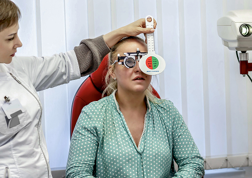
When the patient's condition allows, the main tests are performed on the same day of the appointment. The patient undergoes a pressure measurement, anterior segment assessment, angle examination, and visual function testing. The results are discussed immediately after the examination.
If pupil dilation is unsafe, the doctor chooses a more gentle examination option. Some examinations can be postponed until a follow-up visit after stabilization.
Individualized treatment and monitoring plan
After the examination, the ophthalmologist creates a plan. It may include medications, laser iridotomy, surgery, or follow-up. The patient receives a treatment plan:
- Consultation
- Diagnostics
- Treatment prescription
- Post-Procedure monitoring
If intervention is necessary, we explain the preparation and follow-up appointments. If the patient's condition is stable, the focus is on pressure control and prevention.


FAQ
How is closed-angle glaucoma different from open-angle glaucoma?
Is it possible to cure closed-angle glaucoma without surgery?
What symptoms require urgent attention?
What does the doctor do at the first appointment?
The doctor will review your symptoms, measure your pressure, examine the anterior segment of the eye, and evaluate the angle. Then, if safe, they will check your nerve and visual function. Based on the results, the patient will receive an explanation of the diagnosis and a plan for further action.
Angle-closure glaucoma requires close monitoring because vision depends on prompt diagnosis and appropriate treatment.
Schedule an appointment at K+31: the doctor will evaluate your intraocular pressure, assess your anterior chamber angle, and help you choose a safe plan to preserve your vision.
Clinical Guidelines of the Ministry of Health of the Russian Federation — https://cr.minzdrav.gov.ru/
MSD Manuals, Russian version — https://www.msdmanuals.com/ru/
Russian Medical Journal, Ophthalmology Section — https://www.rmj.ru/articles/oftalmologiya/
CyberLeninka — search for "angle-closure glaucoma" - https://cyberleninka.ru/
CyberLeninka - search on the topic "laser iridotomy glaucoma" - https://cyberleninka.ru/
EyePress - ophthalmological publications - https://eyepress.ru/
Mediasphera - https://www.mediasphera.ru/
Our doctors





















This award is given to clinics with the highest ratings according to user ratings, a large number of requests from this site, and in the absence of critical violations.

This award is given to clinics with the highest ratings according to user ratings. It means that the place is known, loved, and definitely worth visiting.

The ProDoctors portal collected 500 thousand reviews, compiled a rating of doctors based on them and awarded the best. We are proud that our doctors are among those awarded.
Make an appointment at a convenient time on the nearest date
Price
Other services

Appointment to the doctor
Reviews





















Our clinics


Application “Personal Account K+31”












What is angle-closure glaucoma and why is it dangerous?
With this form of the disease, the angle of the anterior chamber narrows or closes. Intraocular fluid must drain through this area, so blocking the outflow leads to increased pressure. The longer the eye remains in this condition, the higher the risk of nerve damage.
Narrow-angle glaucoma is dangerous because symptoms can appear suddenly. Without an examination, it's impossible to determine how closed the angle is or whether there's a threat to vision. Therefore, even brief episodes of fog or pain should be discussed with a doctor.
Why this condition requires prompt medical attention
The doctor's primary goal is to protect the optic nerve. High pressure can damage its fibers, and the patient often doesn't notice the first changes. Pain, fog, and sudden vision loss require urgent examination.