
Megalocornea (large cornea): what it is, how it's diagnosed, and what to do next
When we see a patient with unusually large, expressive eyes, it always warrants special professional attention. Sometimes, such a beautiful appearance conceals megalocornea—a rare condition that requires exceptional vigilance from the doctor and a conscious approach to health from the patient.
At the K+31 clinic, we often encounter people confusing this anomaly with dangerous diseases or, conversely, not paying it due attention.
We explain how megalocornea is diagnosed and why regular ophthalmological follow-up is so important.

specialists

equipment

treatment






Why does iris melanoma develop? Risk factors

Heredity and pigmentation characteristics
Modern medical science cannot yet identify a single, definitive cause that triggers this pathological process. However, long-term clinical observations have identified a group of factors that significantly increase the risk of developing this tumor. A person's natural pigmentation plays a significant role, as statistics show that people with fair skin and blue, gray, or green eyes are significantly more likely to experience this condition. Genetic predisposition and a strong family history are also considered by doctors.
UV radiation, trauma, and other possible triggers
Chronic exposure to sunlight without protective lenses can damage the DNA of iris cells and provoke their malignant transformation. Ophthalmic oncology also associates possible triggers with previous severe eye injuries or chronic inflammatory processes in the anterior segment. Sometimes the trigger is a simple pigmented lesion that existed for many years as a normal birthmark, but under the influence of external factors, transformed into a malignant nodule.
Why does megalocornea develop?
Congenital causes
This condition is based on a genetic factor. It is a classic congenital corneal anomaly that develops in the earliest stages of embryonic development, approximately in the third month of pregnancy.
During this period, for some reason, the regulation of growth of the anterior segment of the eye is disrupted, and the cornea begins to grow faster than other structures.
Hereditary and syndromic forms
In almost 90% of all reported cases, this condition is inherited. X-linked recessive inheritance is most common. This means that it predominantly affects men, while women are carriers of the defective gene. Therefore, when a patient comes to us with a suspected diagnosis, we always conduct a thorough family history and ask whether their father, grandfather, or maternal uncle had large eyes.
When megalocornea is combined with other disorders
Sometimes this pathology is isolated, meaning that other than the size of the cornea, nothing else distinguishes the person from the norm. However, in some cases, there are associated developmental anomalies. A large anterior segment can be part of systemic genetic diseases such as Marfan syndrome, Down syndrome, or Alport syndrome.
In such situations, we are dealing with changes not only in the eyes, but also in the connective tissue, musculoskeletal system, or cardiovascular system.
How to suspect megalocornea

Symptoms in children
Megalocornea in children is usually detected immediately after birth or in the first months of life. Parents or a pediatrician notice that the baby's eyes are unusually large and beautiful. If this is an isolated feature, the child is otherwise undisturbed. The eyes remain calm, there is no redness, and the child reacts normally to light and follows toys.
Symptoms in adults
If not diagnosed in childhood, megalocornea in adults is most often an incidental finding during a routine examination. Patients themselves may not present any specific complaints other than a gradual deterioration in vision. The large anterior chamber of the eye often leads to a shift in focus, leading to associated refractive changes.

When is an urgent visit to an ophthalmologist needed?
A large cornea itself doesn't hurt or cause discomfort. However, there are critical symptoms that require a visit to the doctor without delay. You or your child need an urgent ophthalmological examination if the following signs appear:
- Severe clouding or swelling of the cornea, causing the eye to lose its luster.
- Constant profuse lacrimation and photophobia.
- The child frequently rubs their eyes and is fussy.
- An adult develops eye pain or a feeling of heaviness.
- Vision has sharply deteriorated, and rainbow-colored rings are visible around light sources.
How we diagnose
Slit lamp examination
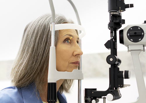
A complete diagnosis of megalocornea is impossible at home; specialized equipment is essential. First, we seat the patient at an instrument called a slit lamp. This is a special microscope that allows us to examine the anterior segment of the eye in detail, under high magnification. We assess the transparency of the cornea, the depth of the anterior chamber, and the condition of the lens and iris to rule out other congenital defects.
Keratometry, pachymetry, and biometry
To accurately confirm the diagnosis, we perform a range of measurement procedures:
- Keratometry — this method allows us to measure the radius of curvature and the true diameter of the cornea with an accuracy of hundredths of a millimeter.
- Pachymetry — ultrasound or optical measurement of the thickness of the cornea. With true megalocornea, tissue thickness remains within normal limits, unlike in other conditions.
- Ocular biometry is an ultrasound examination that helps measure the overall length of the eyeball, the depth of the anterior chamber, and the size of the crystalline lens.
Vision and intraocular pressure testing
We always examine the overall functioning of the visual system. The doctor tests visual acuity and determines the patient's refraction. Measuring intraocular pressure is also a key step. This is critical, as elevated intraocular pressure can completely destroy the optic nerve.
Differential Diagnosis with Glaucoma, Keratoglobus, and Buphthalmos
Our team's primary goal is to accurately differentiate between similar pathologies. We require a detailed differential diagnosis, as a large cornea can mask life-threatening conditions. First and foremost, we rule out congenital glaucoma. In this condition, the eye also enlarges, but this occurs due to critical fluid pressure.
We also distinguish megalocornea from pathologies such as keratoglobus (a spherical protrusion and thinning of the cornea) and buphthalmos (a generalized stretching of the entire eyeball in advanced glaucoma).
| Signature | Megalocornea | Congenital glaucoma | Keratoglobus |
|---|---|---|---|
| Corneal size | Enlarged | May be enlarged | Enlarged |
| Intraocular pressure | Usually normal | Often elevated | Usually Normal |
| Corneal condition | Usually clear | Possible swelling/cloudiness | Thinning, bulging |
| Tactics | Observation | Treatment as indicated | Individually determined |
General information
What are the dangers of megalocornea and what are the risks?
Association with increased intraocular pressure
Although in the isolated form, intraocular pressure is initially within normal limits, the anatomical features of the eye create risks for its future increase. Due to the excess space in the anterior chamber, the ligaments that hold the lens in place can stretch and weaken. If the lens becomes displaced, it can block the outflow of aqueous humor.
Potential problems with refraction and vision quality
A large optical window is often accompanied by changes in light refraction. Such patients often have high myopia (nearsightedness) or complex astigmatism. Without proper correction, these conditions interfere with the formation of a clear image on the retina, which in children can lead to amblyopia (lazy eye syndrome).
When additional monitoring is required
Additional and more frequent monitoring by an ophthalmologist is necessary if we observe concomitant changes in the structure of the eye. Weakening of the lens ligament apparatus, early development of cataracts, or peripheral retinal thinning are all reasons to visit the clinic more frequently than usual.


FAQ
What is megalocornea and does it need treatment?
Can megalocornea affect vision?
How to distinguish megalocornea from congenital glaucoma?
What tests are needed if a diagnosis is suspected?
What to do after the diagnosis is confirmed
If treatment is not required
If all the test results confirm an isolated form of the anomaly, you simply need to accept this individual characteristic of your body. Glasses or lenses will help correct the accompanying myopic focus, and you will be able to lead a completely full, active life without any strict limitations.
When is a follow-up visit needed?
Your doctor will schedule your next examination. Typically, if the results are stable, we expect you to see them in 6 or 12 months. The main thing is to stick to this schedule and not miss appointments, even if you think your eyes are perfectly fine.
How to schedule an examination
If you notice that your child has unusually large corneas, or your doctor has expressed similar suspicions to you during a routine examination, do not delay your appointment with a specialist. Schedule an appointment at the K+31 clinic by phone or using the form on our website. We will conduct an expert examination, dispel all your doubts, and help you maintain your eye health for years to come.

Clinical Guidelines of the Ministry of Health of the Russian Federation — https://cr.minzdrav.gov.ru/
MSD Manuals, Russian version, ophthalmology section — https://www.msdmanuals.com/ru/professional/ophthalmology
Russian Medical Journal, ophthalmology section — https://www.rmj.ru/articles/oftalmologiya/
Bulletin of Ophthalmology (MediaSphera) - https://www.mediasphera.ru/journal/vestnik-oftalmologii
EyePress - https://eyepress.ru/
CyberLeninka - https://cyberleninka.ru/
Our doctors





















This award is given to clinics with the highest ratings according to user ratings, a large number of requests from this site, and in the absence of critical violations.

This award is given to clinics with the highest ratings according to user ratings. It means that the place is known, loved, and definitely worth visiting.

The ProDoctors portal collected 500 thousand reviews, compiled a rating of doctors based on them and awarded the best. We are proud that our doctors are among those awarded.
Make an appointment at a convenient time on the nearest date
Price
Other services

Appointment to the doctor
Reviews





















Our clinics


Application “Personal Account K+31”












What is megalocornea?
Definition in simple terms
In its simplest form, it is a congenital anomaly of the cornea in which its horizontal diameter significantly exceeds the average norm. The transparent "eye window" increases in size even before birth, but the corneal tissue itself retains its normal structure, transparency, and thickness.
How megalocornea differs from the normal diameter
In most healthy people, the cornea's diameter is approximately 11–12 millimeters. A person has a large cornea when this diameter exceeds 12 millimeters in a newborn baby and exceeds 13 millimeters in an adult.
Our experience shows that isolating this feature in itself does not prevent the eye from refracting light correctly, but it does create specific anatomical conditions that we must control.