
Tumors and growths of the eyelid skin: how to recognize them and when to remove them
Neoplasms on the skin of the eyelids are common, and most are harmless. But it's especially important to quickly distinguish between a benign growth and something that requires medical attention in the eyelid area. The skin here is thin and the eye is close, so self-medication is dangerous.
We'll explain the different types of eyelid neoplasms, the warning signs, and why histology is necessary.

specialists

equipment

treatment






What types of eyelid neoplasms are there?
Eyelid lesions are usually divided into several groups based on their nature. The nature of the lesions determines the appropriate tactics: observation, removal, or further examination. Let's look at the main types.
Benign tumors
Benign growths are the most common. These include eyelid papillomas, eyelid moles, cysts, warts, and keratomas. They grow slowly, are usually painless, and do not change for a long time.
These growths are most often a concern as a cosmetic defect or when they catch on eyelashes or clothing. However, even a benign growth should be examined by a doctor if it begins to change. The external similarity of different growths can be deceptive.
Pre-cancerous changes and suspicious elements
A separate group are changes that are not tumors in themselves, but require monitoring. They can degenerate over time, so they are monitored especially closely. Elements that grow rapidly, bleed, or are unevenly colored are considered suspicious.
The doctor monitors such changes on a schedule and, if necessary, refers the patient for additional examination. This helps detect dangerous degeneration at an early stage. Therefore, it's important not to skip follow-up examinations.
Malignant tumors
Malignant tumors are less common on the eyelids. They are dangerous because they invade surrounding tissue and spread without treatment. A persistent ulcer, loss of eyelashes in the area, and a dense, growing nodule should raise concern.
These tumors require prompt diagnosis and mandatory laboratory confirmation. The earlier they are detected, the higher the chance of careful and complete removal. Therefore, it's best to have any persistent, unusual growth examined by a doctor.
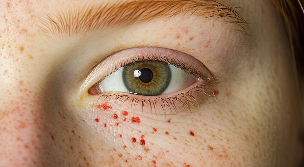
Why do growths appear on the eyelids?
It's difficult to pinpoint the exact cause of each condition, but certain factors are known to increase the risk. The main one is prolonged sun exposure to the thin skin of the eyelids. Age, heredity, and skin characteristics also play a role.
Chronic inflammation of the eyelid margins, viral infections, and decreased immunity also contribute. Sometimes growths appear at the site of old injuries and scars. Therefore, if any persistent changes in the skin of the eyelids occur, it's wise to consult a doctor.
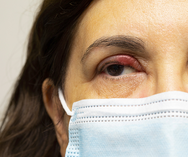
What symptoms should you be concerned about?
Most growths are benign, but there are signs that require examination. These indicate that the growth is changing or interfering with the eye. Let's look at them in more detail.
Rapid growth, bleeding, color change
It's alarming when a growth rapidly grows, bleeds, or crusts over. Changes in color, shape, and unclear, blurry borders are all warning signs. These signs are a reason to see a doctor immediately.
Itching, pain, inflammation, and irritation when blinking
Itching, pain, and persistent inflammation in the area of the growth should be a warning sign. If the growth clings to the eye and is injured when blinking, it becomes constantly irritated. This condition interferes with vision and increases the risk of complications.
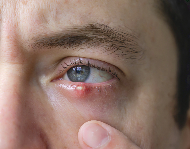
A formation at the ciliary margin or on the moving part of the eyelid
Particular attention should be paid to lesions at the cilia margin and on the moving part of the eyelid. They interfere with blinking more quickly here and can affect the cornea. Removing anything in such a delicate area on your own is unacceptable.
To make it easier to understand, below are the signs that indicate a need to see a doctor:
- Rapidly increasing in size
- Bleeding or crusting
- Changes color, shape, or borders
- Interferes with blinking or catches on eyelashes
- Frequently becomes inflamed or injured
If you have any of these signs, don't wait: it's best to see a specialist and determine the nature of the lesion.
How is the diagnosis carried out?
The doctor's main task is to understand the nature of the growth and decide whether it needs to be removed. In the delicate eyelid area, external appearance alone cannot be relied upon. Let's review the examination steps.
Examination by an ophthalmologist or dermato-oncologist
First, an ophthalmologist or dermato-oncologist examines the lesion and assesses its size, shape, and location. The doctor checks to see if the lesion is affecting the cornea or eyelashes. At this stage, an initial understanding of its nature is already formed.
Dermatoscopy and photofixation
For a detailed examination, dermatoscopy—examining the affected area under magnification—is used. This method helps visualize structures and vessels that are not visible to the naked eye. Photographic documentation allows for dynamic comparison of the condition during follow-up visits.
When is a biopsy and histology needed?
If a tumor appears suspicious, it is sent for histology after removal. Laboratory testing accurately determines the nature of the tissue and rules out dangerous changes. Sometimes a biopsy is performed beforehand to refine the treatment plan.
Histology is especially important when the tumor is atypical or rapidly changing. It removes any doubts and confirms that the tumor has been completely removed. Therefore, we never neglect this step.
Methods for removing skin growths on the eyelids
| Method | When is it suitable? | Advantages | Important considerations |
|---|---|---|---|
| Surgical excision | Suspicious or large lesions | Complete removal, suitable for histology | Requires careful technique |
| Radiowave ablation | Small superficial lesions | Precision, gentle treatment | The decision is made by the doctor |
| Laser removal | Some benign lesions | Quick and accurate | Not always suitable if histology is required |
Surgical excision
Surgical excision is used for suspicious and large lesions. This method allows for the removal of the entire lesion and sending the tissue for histology. This is especially important when dangerous changes must be accurately excluded.
With this type of removal, the surgeon also removes a small area of healthy tissue at the edge, if necessary. This reduces the risk of part of the lesion remaining. A histological examination provides the final conclusion.
Radio wave removal
Radio wave removal is suitable for small superficial lesions. It is targeted and gentle on the surrounding skin. This method produces a precise result in the delicate eyelid area.
Laser removal
Laser removal is used for some benign lesions. It is quick and gentle, but not always suitable. If histological examination is required, the doctor will choose another method.
How the doctor chooses the method in each case
The method is selected individually, for each specific case. The doctor takes into account the nature of the element, its size, location, and the need for laboratory testing. Therefore, diagnostics are always performed before the procedure.
General information
Advantages of professional eyelid removal
The eyelids are a delicate and mobile area near the eye, where precision and experience are essential. Professional removal reduces risks and preserves appearance. Below is a summary of the benefits for the patient.
Accurate diagnosis before the procedure
Before removal, we determine the nature of the lesion. This helps us choose the right method and avoid missing dangerous changes. The decision is made consciously, based on the diagnostic results.
A gentle approach and maintained aesthetics
In the eyelid area, it is important to preserve both function and appearance. We work carefully to minimize scarring. Our meticulous technique helps preserve the natural shape of the eyelid.
Result monitoring and post-removal follow-up
After the procedure, we monitor the skin's healing. We provide clear care recommendations and, if necessary, schedule a follow-up visit. If indicated, we send the specimen for histology.
FAQ
Short answers to the questions most often asked during an appointment
How can you tell if a growth on your eyelid is dangerous?
Should any growth on the eyelid be removed?
Is eyelid growth removal painful?
Why is histology necessary after removal?

Clinical Guidelines of the Ministry of Health of the Russian Federation
Bulletin of Dermatology and Venereology
Russian Medical Journal — ophthalmology
Russian Medical Journal - dermatology
CyberLeninka - search on the topic “tumors” century"
CyberLeninka - search on the topic “new growths of the eyelids”
Our doctors





















This award is given to clinics with the highest ratings according to user ratings, a large number of requests from this site, and in the absence of critical violations.

This award is given to clinics with the highest ratings according to user ratings. It means that the place is known, loved, and definitely worth visiting.

The ProDoctors portal collected 500 thousand reviews, compiled a rating of doctors based on them and awarded the best. We are proud that our doctors are among those awarded.
Make an appointment at a convenient time on the nearest date
Price
Other services

Appointment to the doctor
Reviews





















Our clinics


Application “Personal Account K+31”












What are new growths on the skin of the eyelids?
A neoplasm is an overgrowth of skin cells or its structures. Various elements can appear on the eyelids:
This area is particularly sensitive. It is located near the eyelashes, mucous membranes, and the eye itself, so even a benign growth can interfere with blinking, irritate the surface of the eye, and affect vision.
Most of these elements grow slowly and do not cause significant symptoms for a long time. However, observation is necessary if the growth enlarges, changes color, bleeds, becomes inflamed, or reappears after removal.
In such cases, the doctor assesses the risk, rules out recurrence, and decides whether further diagnostics are necessary. Understanding the nature of an element based solely on its appearance isn't always possible, and that's the main difficulty.