Sectoral retinal laser coagulation in Moscow
A tear, thinning, or dystrophy of the peripheral fundus may not affect vision for a long time. The patient lives a normal life and is unaware of the risk zone.
During the consultation, we assess not only visual acuity, but also the condition of the fundus, vitreous, and macula. If indicated, retinal laser coagulation in Moscow helps to specifically strengthen the affected area.

specialists

equipment

treatment


What is sectoral laser coagulation of the retina?
This method involves targeted laser treatment of a limited area of the fundus. The laser forms coagulates—small areas of tissue adhesion around the defect or thinning.
This helps the doctor isolate the problem area. The decision is made after an in-person diagnosis.
How does the sectoral technique differ from the barrier and panretinal methods?
The sectoral method is chosen for localized changes. Barrier laser coagulation, or barrier coagulation, helps isolate the tear or edge of a localized detachment. Panretinal treatment is used for other purposes, such as ischemia due to diabetic retinopathy. The extent of the procedure depends on the diagnosis.
Before treatment, we explain the purpose of each method.
| Procedure Type | When Applied | Volume of Treatment | Purpose |
|---|---|---|---|
| Sectoral | Localized Dystrophy, Rupture, Thinning | Single Area | Strengthen Risk Area |
| Barrier | Rupture or Localized Detachment Along the Edge | Along the Defect | Limit the spread of the process |
| Panretinal | Diabetic retinopathy, ischemia | Large area, excluding the macula | Reduce the impact of ischemia |
The doctor selects a specific method after examination, imaging, and risk assessment.
What are the results of the procedure?
Laser coagulation of the retina does not reverse the structure of damaged tissue. The doctor uses a laser to create adhesion points around the damaged area to reduce the risk of widening the tear or worsening the thinning. After the procedure, the condition of the fundus is monitored dynamically.
When do we recommend retinal laser coagulation?

We don't prescribe laser treatment based solely on complaints. Sometimes vision remains clear, but there are already dangerous changes in the periphery. The opposite can also happen: symptoms are pronounced, but the cause is:
- In the vitreous body
- Macula
- Optic nerve or inflammation
Therefore, indications for laser photocoagulation are determined after an examination.
Retinal tears and thinning
This is a tissue defect through which fluid can leak under the neurosensory layer. The risk is higher:
- With recent flares
- Trauma
- High myopia
- A similar problem in the second eye
In these cases, sectoral laser coagulation allows for localized treatment.

Peripheral Retinal Dystrophies
This clinical case presents with changes in areas of the retina closer to the fundus. The risk depends on the type of dystrophy, the depth of thinning, myopia, and the connection with the vitreous. If there are dangerous signs, the doctor may recommend laser retinal strengthening.
Risk of Retinal Detachment
This clinical case requires prompt medical evaluation. If retinal detachment has already begun, surgical treatment may be necessary. If changes are limited, the doctor sometimes considers laser treatment as a preventative measure. The periphery is often visible only after pupil dilation.
We most often discuss laser with patients in five situations:
- Peripheral retinal dystrophy with dangerous features
- Local retinal tear
- Peripheral thinning with traction
- Prevention of retinal detachment in high-risk patients
- Follow-up after a retinologist's examination
After the list, the doctor returns to the examination data: the size of the zone, location, and condition of the other eye are important.
General information
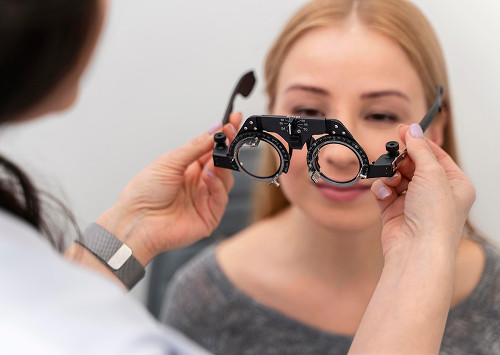
How is sectoral laser coagulation performed?
The procedure is explained to the patient in advance. Treatment is typically performed on an outpatient basis, without hospitalization. Before beginning, the doctor reviews the examination data, checks on the patient's well-being, and warns about possible sensations.
Preparation for the Procedure
Preparation for the procedure includes pupil dilation, eye drops, and insertion of a contact lens, if needed for focusing. There is usually no significant pain. Possible side effects include:
- Pressure from the lens
- Bright flashes
- Short-term discomfort
The doctor asks the patient to look in a designated direction.
Laser Treatment Stages
During the session, pulses are applied according to the planned schedule. Laser coagulation of the retina creates a series of coagulates around the risk zone or along the edge of the defect.
Parameters are selected individually, taking into account pigmentation, media transparency, site location, and tissue response. This type of laser treatment of the retina is not performed according to a one-size-fits-all procedure.
Is it painful and how long does the session last?
Most patients tolerate the session well. Discomfort is associated with bright light, contact lenses, or the treatment of specific areas.
The duration depends on the treatment area and access to the area. We explain the procedure in advance to reduce anxiety.


Frequently Asked Questions
What is sectoral laser coagulation of the retina?
Is sectoral laser coagulation painful?
When does a doctor recommend this procedure?
What is recovery like after the procedure?

Our doctors


This award is given to clinics with the highest ratings according to user ratings, a large number of requests from this site, and in the absence of critical violations.

This award is given to clinics with the highest ratings according to user ratings. It means that the place is known, loved, and definitely worth visiting.

The ProDoctors portal collected 500 thousand reviews, compiled a rating of doctors based on them and awarded the best. We are proud that our doctors are among those awarded.
Make an appointment at a convenient time on the nearest date
Price
Other services

Appointment to the doctor
Reviews


Our clinics

Application “Personal Account K+31”


How we conduct diagnostics before the procedure
Before laser treatment, an accurate fundus map is required. We do not perform laser retinal treatment based on an old report or symptom description. First, the doctor clarifies:
Fundus examination with pupil dilation
Pupil dilation is necessary to examine the periphery of the fundus. Without drops, the doctor may not see areas where thinning, dystrophy, or tears are more likely to form.
After instillation, near vision may become temporarily blurred, and light may appear too bright. It is best not to drive on the day of the examination. An ophthalmologist performs the examination.
OCT, ophthalmoscopy, fundus camera
Ophthalmoscopy allows the doctor to directly examine the fundus. This allows for the evaluation of the vessels, optic disc, periphery, and areas of potential thinning. Retinal OCT is used to examine the macula and the layered structure of the tissues in detail. A fundus camera saves the image for comparison at subsequent visits. If the patient's picture is complex, a retinologist also evaluates the patient.
Why is it important to assess risks before laser treatment?
The laser must be precise. Insufficient volume may not solve the problem, and unnecessary exposure is unnecessary. We evaluate the transparency of the media, inflammation, the location of the defect, and access to the desired area. Contraindications are also discussed at this stage.