Treatment of Humerus Fractures in Children
Children lead active lifestyles, so upper limb injuries are frequently diagnosed by surgeons. Professional treatment of a humerus fracture requires prompt medical diagnosis and a careful approach. Orthopedists always consider the anatomical features of the growing skeleton when choosing a treatment strategy. The speed of healing and full restoration of the arm depend on the competent intervention of specialists.

specialists

equipment

treatment


Symptoms of a humerus fracture

A serious injury can be recognized by a number of specific signs that appear immediately. Classic symptoms of a humerus fracture include the patient's acute reaction to any contact with the limb. The severity of clinical manifestations always depends on the severity of the injury.
Pain, swelling, limited motion
At the moment of mechanical impact, the patient experiences sharp, unbearable pain. A short time later, increasing pain and swelling of the soft tissues in the area of the defect occur. The victim is physically unable to raise the arm or bend the elbow due to muscle spasm. Any attempt to simply move the fingers causes severe physical discomfort.

Arm Deformation and Forced Position
With extensive tissue damage, the anatomical shape of the limb is visually altered. The arm begins to appear shortened or bent at an unnatural angle.
The child reflexively presses the affected limb against their body, supporting it with the healthy hand. This forced position helps to slightly reduce the intensity of the pain.
When to See a Doctor Immediately
Any injury to the upper limb in children requires a qualified medical evaluation. An urgent visit to the doctor is necessary if the fingers turn pale or if there is complete loss of skin sensation.
A cold hand and a bluish tint directly indicate damage to major blood vessels. Open wounds are also an immediate reason to call an ambulance if a child has a complex shoulder fracture.

A reliable determination of the nature of the injury allows the specialist to select the optimal treatment. The standard medical examination includes a thorough visual examination and the mandatory use of instrumental techniques. The doctor also rules out possible hidden vascular or nerve ruptures.
After the initial examination, the orthopedic traumatologist checks not only the injury site but also the function of the hand:
- Sensation
- Circulation
- Response to gentle movements.
X-rays provide the primary confirmation of the diagnosis, as they show the location of the fragments and the extent of the injury. In complex cases, CT or MRI scans are added to the examination if a more detailed assessment of the joint, ligaments, or deep tissues is needed.
General information
Treatment Methods for Humerus Fractures in Children
The choice of treatment strategy is based on detailed examination results. Conservative or surgical treatment of humerus fractures in children is aimed at ideal tissue fusion. Doctors prefer the most gentle and safe medical techniques.
Immobilization and Conservative Treatment
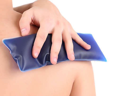
For uncomplicated injuries, it is sufficient to securely immobilize the limb in a static position. For this purpose, medical immobilization using modern polymeric materials is used. A properly applied plaster cast or orthosis limits movement for three to six weeks. Under complete rest, the body naturally forms a dense callus.
Reposition and Fixation in Case of Dislocation
If severe divergence of the edges is diagnosed, the tissues must be returned to their natural anatomical position. This medical procedure is called repositioning, and it is always performed under effective anesthesia.
After the fragments are aligned, a plaster cast is applied, and then a repeat x-ray is taken. The specialist must ensure that the bone axis is completely aligned.
When Surgery Might Be Necessary
Surgery becomes necessary in the presence of multiple or unstable fragments. Fracture surgery involves the insertion of special metal pins, screws, or titanium plates.
This method is called osteosynthesis; it ensures rigid internal support of the problem area. Metal structures are usually removed after confirmed tissue healing.
Before approving a treatment plan, the doctor analyzes the entire range of clinical indicators.
Below is a comparative table demonstrating key approaches to providing specialized care. The final decision on the use of a specific technique is made exclusively by a specialized surgeon.
| Method | When used | Advantages | Limitations |
|---|---|---|---|
| Immobilization | For injuries without displacement | A gentle approach, ideal for young patients | Requires systematic monitoring by a physician |
| Reposition | For severe displacement of fragments | Helps to accurately restore the correct position | A repeat x-ray is required |
| Surgery | For unstable or Complex injuries | Ensures the most precise fixation | Involves a more complex recovery phase |


Frequently Asked Questions
How can I tell if my child has a humerus fracture?
How long does it take for a humerus fracture to heal in children?
Is surgery always necessary if a humerus fracture is diagnosed?
When is it permissible to return to normal activities and sports?
Such permission is granted only after full range of motion has been restored and healing has been confirmed. Exercise intensity is increased very slowly to reliably rule out the risk of re-injury.
Treatment strategies for a humerus fracture depend on the imaging, the child's age, and the condition of the arm after the injury. X-rays are taken first, then the doctor evaluates for signs of vascular or neurological disorders and, if necessary, orders follow-up imaging. As the traumatologist notes, "If a child's humerus fracture is detected early, properly stabilized, and the healing process is monitored, the chances of a good healing are high." After the initial treatment, physical therapy may be added to restore arm motion without putting unnecessary strain on the arm.


This award is given to clinics with the highest ratings according to user ratings, a large number of requests from this site, and in the absence of critical violations.

This award is given to clinics with the highest ratings according to user ratings. It means that the place is known, loved, and definitely worth visiting.

The ProDoctors portal collected 500 thousand reviews, compiled a rating of doctors based on them and awarded the best. We are proud that our doctors are among those awarded.
Make an appointment at a convenient time on the nearest date
Price
Other Services

Appointment to the doctor


Our clinics

Application “Personal Account K+31”


What is a humerus fracture in a child?
A bone fracture in the area from the shoulder joint to the elbow completely disrupts the weight-bearing function of the upper limb. A similar fracture of the humerus in a child occurs as a result of a strong blow, a fall on an outstretched arm, or a sharp twist. Anatomically, this tubular bone is divided into upper, middle, and lower parts. The precise location of the defect directly determines the subsequent medical plan.
Types of fractures: non-displaced, displaced, closed, open
These injuries are differentiated by the position of the bone and the condition of the skin. With a non-displaced fracture, the limb axis remains normal. With a displaced fracture, the bone fragments shift and no longer hold their normal position. A closed fracture does not break the skin. An open fracture is immediately noticeable: a wound appears, often with bleeding.
Why are fractures in children different from those in adults?
The musculoskeletal system in children is covered by a dense and elastic membrane called the periosteum. It prevents fragments from separating too much, creating a characteristic "greenstick" effect. Young patients also have exposed cartilage growth zones. Injuries to these sensitive areas require increased medical attention to prevent future arm shortening.