Zygomatic bone fracture in children: symptoms, diagnosis, treatment, and recovery
Zygomatic bone fractures are the second most common facial bone injuries after nasal injuries. This is a very complex injury, as the zygomatic bone connects the frontal, temporal, and maxillary bones.
Treating a zygomatic bone fracture in children not only preserves the child's appearance but also ensures proper visual and dental function.

specialists

equipment

treatment


Causes of Trauma in Children
The causes of maxillofacial injuries evolve as a child ages, moving from everyday carelessness to injuries sustained in a fight.
Falls and household blows
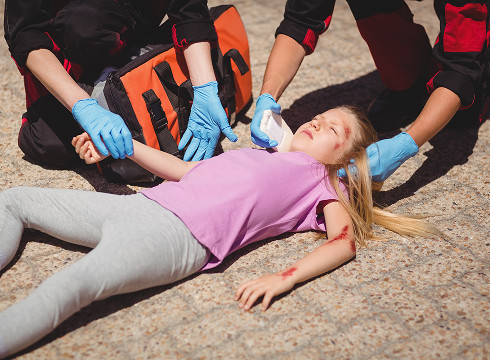
For younger children (under 7 years old), the main factor is poor motor coordination. Falling from a crib, high chair, or hitting an unprotected corner of furniture often causes a child's cheekbone fracture. In everyday life, injuries often occur as a direct blow, with all the energy impacting the protruding part of the cheekbone.
Sports injuries
Between the ages of 8 and 14, organized and spontaneous sports activities become more common. Maxillofacial trauma is common in hockey, martial arts, and football (elbow or ball strikes). The danger of sports injuries is that they often occur at high speeds, increasing the risk of bone fragment displacement. Even with protection, a sharp side impact can cause a zygomatic arch fracture.
Road traffic and severe impact injuries
Road accidents are the most severe category of injuries. Even with child safety seats, sudden braking or airbag deployment can cause a blow to a child's face. In these cases, a child's zygomatic bone fracture is often accompanied by a traumatic brain injury, jaw fractures, and soft tissue damage. Such combined injuries require immediate intervention by a surgical team.
Symptoms of a zygomatic bone fracture

Clinical signs can vary from subtle to pronounced, depending on the impact vector and the patient's age.
Pain, Swelling, and Hematoma
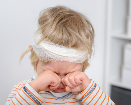
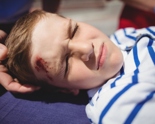
Swelling of the cheek appears immediately. It quickly spreads to the eyelids. The eye may become red, and a periorbital hematoma appears on the lower eyelid.
The child experiences pain, which intensifies when attempting to speak or change facial expressions. They may also complain of a throbbing in the cheekbone area.
Facial Asymmetry and Deformity
A characteristic symptom is a "flattening" of the cheekbone on the affected side. When looking at the child's face from above, tissue depression is noticeable. However, it is important to remember that after 1-2 hours, increasing swelling can completely obscure this symptom, creating a false appearance of well-being. Therefore, facial asymmetry should be assessed dynamically.

Limited Mouth Opening and Chewing
The displaced bone presses against the lower jaw, preventing the child from opening the mouth normally. A little later, contracture spasm of the masticatory muscles develops, preventing the child from chewing even soft foods.
The pain when chewing becomes unbearable. Any attempt to open the mouth wide is accompanied by a characteristic clicking sound or locking of the joint.
Numbness, malocclusion, and visual complaints
Damage to the infraorbital nerve causes numbness in the cheek and upper lip. This frightens the child, who no longer feels their face.
If the inferior orbital wall is affected, double vision (diplopia) and enophthalmos (sunken eye) occur.
An abnormal bite is another consequence of trauma. Because the teeth don't close properly, the upper jaw becomes deformed.
When Urgent Help Is Needed
Parents often underestimate the severity of a blow to the face, dismissing it as a simple bruise. However, there are conditions that require immediate medical attention.
Warning signs that require more attention
Contact a doctor immediately if you notice any of the following symptoms in your child:
- Double vision: the child sees two identical objects instead of one.
- They have no feeling in their upper jaw.
- The pupils are different sizes, and the eye moves poorly.
- Bleeding from the nose or mouth.
- Numbness in the cheek that does not go away within an hour after the blow.
- The mouth opens less than the width of one finger.
What to do before the doctor's examination.
Place the child so that the head is elevated (this reduces blood flow and swelling). Apply a cold compress to the cheekbones. If the teeth are damaged or there is bleeding from the mouth, help them rinse with cool water.
What not to do
It is strictly forbidden:
- Trying to "feel" the fracture site with your fingers - this may lead to further displacement.
- Applying heating pads or alcohol compresses.
- Asking the child to clench their jaws tightly to "check their bite."
- Using ointments like "badyagi" or warming gels before a diagnosis is made.
- Allowing the child to actively blow their nose, as this can cause subcutaneous emphysema (inflation of the face with air).
General information
Fracture Diagnosis
Diagnostic work in pediatric maxillofacial surgery should be as quick and atraumatic as possible.
Specialist Examination
A pediatric traumatologist or maxillofacial surgeon begins with palpation of the "bone steps"—uneven areas along the orbital and cheekbone margins. An important step is checking visual function and the sensitivity of the trigeminal nerve innervation zones. The doctor also examines the oral cavity for the integrity of the alveolar process.
X-ray and CT scan of the facial skeleton
X-ray of the facial skeleton in axial and AP projections is a basic diagnostic method that does not reveal all fractures.
Today, doctors recommend CT scans of the facial skeleton with 3D reconstruction. Computed tomography (CT) scans allow us to visualize cracks in the thin orbital walls and assess the condition of the maxillary sinus. This is critical for deciding whether to perform surgery.
Specifics of Child Examination
Considering the patient's age, doctors try to avoid unnecessary stress. If the child is too young to lie still for 5-10 minutes, the CT scan is performed under light sedation. The feasibility of the examination is always assessed to ensure a diagnosis with minimal radiation exposure.


Frequently Asked Questions
Should a child undergo a CT scan if a cheekbone fracture is suspected?
Yes, a CT scan allows us to assess the nature of the injury and determine whether there is any displacement. A regular X-ray does not show minor damage to the orbital floor, which is critical for preserving vision.
Is surgery always necessary?
No. It all depends on whether the eye and jaw function are preserved.
How long does recovery take?
The recovery time depends on the severity of the injury, the child's age, and the treatment method. On average, active recovery takes 3-4 weeks, but full bone maturation takes months.
Conclusion
A zygomatic bone fracture in a child is a dangerous injury. However, X-rays and CT scans of the facial skeleton can help detect the problem early and prescribe the correct treatment.
Any maxillofacial injury should be examined by a doctor. Do not self-medicate; trust your child's health to specialists.


This award is given to clinics with the highest ratings according to user ratings, a large number of requests from this site, and in the absence of critical violations.

This award is given to clinics with the highest ratings according to user ratings. It means that the place is known, loved, and definitely worth visiting.

The ProDoctors portal collected 500 thousand reviews, compiled a rating of doctors based on them and awarded the best. We are proud that our doctors are among those awarded.
Make an appointment at a convenient time on the nearest date
Price
Other Services

Appointment to the doctor


Our clinics

Application “Personal Account K+31”


What is a zygomatic bone fracture?
This is a fracture of the zygomatic bone or its processes. The zygomatic bone forms the lateral portion of the midface, contributing to the floor and outer wall of the orbit, as well as the zygomatic arch. A zygomatic bone fracture is rarely isolated; more often, it involves adjacent structures, creating a so-called zygomatic-orbital fracture.
Characteristics of the Child's Facial Skeleton
The child's facial skeleton is unique. It differs from the adult skeleton in its high degree of vascularization (blood supply). Children's bones heal and fuse more quickly. However, not everything is so rosy: even the slightest impact can cause cheek swelling in children.
Children's bones are more flexible. When struck, they do not break, as in adults, but rather crack (similar to the breaking of a fresh branch). Underdevelopment of the maxillary sinuses in young children makes the zygomatic region more monolithic. A blow of great force is required to fracture it.
What is the danger of injury to the zygomatic-orbital region?
The zygomatic-orbital region is a strategic area. The main danger of a fracture here is the risk of damage to the orbital contents. Displacement of fragments can cause entrapment of the inferior extraocular muscles, leading to diplopia. Furthermore, the zygomaticofacial and zygomaticotemporal nerves pass through the zygomatic bone, and just below, the infraorbital nerve. Trauma to these nerves leads to long-term sensory loss. Another danger is the formation of a bony blockade of the coronoid process of the mandible, which causes persistent limitation of mouth opening.