
Compression fracture of the thoracic spine in children: treatment, diagnosis and recovery
A thoracic compression fracture in children is a diagnosis that almost always leaves parents with one key question: how serious is it? The answer depends on the extent of the injury, the child's age, and how quickly the child seeks medical attention.
A child's spine can recover quickly. However, a good prognosis doesn't come naturally. Diagnosis, weight-bearing restrictions, and specialist observation are necessary. If a thoracic compression fracture is confirmed, treatment is individualized, taking into account pain, imaging, posture, mobility, and the child's overall condition.
In the first few days, it is especially important not to aggravate the injury. Avoid warming the back, massaging the painful area, and forcing the child to actively move through the pain. The doctor then explains how the treatment will proceed and when rehabilitation can begin.

specialists

equipment

treatment






Causes and risk factors in children

Children's activity knows no bounds, and this is a natural stage of development. Unfortunately, it is precisely during the process of exploring the world and testing their physical capabilities that compression fractures of the thoracic vertebra most often occur. Situations leading to injury are usually associated with sudden compression of the spinal column along its vertical axis.
Falls and household injuries
Falls from height are the leading cause. This could include falling from a swing, a tree, a garage, or a high wall bar at a playground. The most dangerous landings are considered to be on straight legs, on the buttocks, or falling backwards onto the back. At this moment, the kinetic energy of the impact instantly passes through the pelvis or legs to the spinal column, causing its sudden compression.

Sports, jumping, and axial loading
Sports injuries are the second most common cause. Children involved in gymnastics, acrobatics, parkour, equestrian sports, or diving are particularly at risk. Trampoline centers, popular today, pose a significant risk. An awkward landing on the trampoline net or a mid-air collision with another child creates enormous stress on the thoracic spine.
In such cases, a compression fracture of the spine in children can occur even with a relatively low jump if the force vector coincides with spinal flexion.
When the risk of injury is higher
There are factors that make bones more vulnerable. These include periods of active growth spurts, when the muscular corset cannot keep up with the elongation of the skeleton, leading to postural abnormalities. A lack of calcium and vitamin D, as well as a sedentary lifestyle that weakens the back muscles, significantly reduce the strength of the vertebrae.
Under these conditions, even a slight fall from standing height can cause injury.
Symptoms to look out for
Recognizing the problem visually can be extremely difficult, as symptoms in children are often vague. However, there are clear markers that, if noticed, should prompt parents to seek immediate medical attention.
Back pain and limited mobility
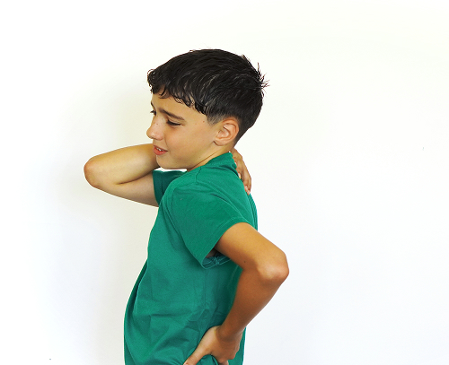
The main symptom is back pain in a child that occurs at the moment of impact. It can be sharp, shooting, or, conversely, dull and aching.
Indirect signs in a child
Quite often, symptoms of a thoracic compression fracture include a brief pause in breathing—post-traumatic apnea. The child may report feeling "breathless" or unable to breathe for several seconds after the fall. Tension in the back muscles around the site of the injury may also be observed.
How is the diagnosis performed?
Since self-diagnosis of back injuries is absolutely unacceptable, professional diagnosis of a compression fracture is crucial. This should be comprehensive and performed in a well-equipped medical facility.
Examination by a pediatric traumatologist-orthopedist
The first step is always a clinical examination. A competent pediatric traumatologist-orthopedist carefully collects the patient's medical history:
- How exactly did the child fall?
- From what height?
- What are the child's complaints?
The doctor palpates the spinous processes of the vertebrae, checks reflexes, sensation, and muscle strength in the legs. Specific stress tests can help determine the location of underlying pain.
Chest X-ray
A basic and mandatory imaging method is a thoracic spine X-ray, which is performed in two projections—AP and lateral. These images allow the doctor to assess the shape of the vertebrae, identify a decrease in the height of their anterior sections, and confirm wedge-shaped deformity. An X-ray provides the first objective picture of the underlying condition.
CT and MRI: When are they needed?
If X-rays don't provide definitive answers or the injury appears complex, more accurate methods are used. A CT scan of the spine is prescribed for a detailed 3D examination of the bone structure to rule out hidden cracks and small fragments. An MRI of the spine, on the other hand, is indispensable for assessing the condition of soft tissues—intervertebral discs, ligaments, blood vessels, and the spinal cord.
To clearly illustrate the differences between the methods, we have prepared a comparison table.
| Method | What it shows | When it is used | Limitations |
|---|---|---|---|
| X-ray | Vertebral shape, vertebral body height | Primary diagnosis | Doesn't always detect hidden changes |
| CT scan | Detailed bone structure | In controversial cases | Radiation exposure |
| MRI | Soft tissues, ligaments, spinal cord | If complications are suspected | Takes longer |
By analyzing the data in this table, the doctor develops the safest and most informative examination strategy, avoiding unnecessary radiation exposure to the growing child, while not missing any important details of the injury.

Treatment of thoracic compression fractures in children
Once the diagnosis is confirmed, targeted therapy begins. Evidence-based medicine dictates that treatment of a compression fracture in a child should be strictly individualized. It depends on the severity of the thoracic compression fracture, the patient's age, and the presence of concomitant neurological symptoms.
Conservative treatment
In the vast majority of cases, if the injury is stable and the spinal cord is intact, a conservative approach is used. The main goal of this stage is to relieve the spinal column, relieve pain, and create ideal conditions for bone fusion. Proper treatment for a thoracic compression fracture allows the vertebral body to restore its density and, as the child continues to grow, partially or completely return to its normal shape.
Immobilization, regimen, and pain management
The basis of conservative therapy is strict bed rest and immobilization on a rigid support. The child is prohibited from sitting or standing. Pain medications are prescribed to relieve spasms and discomfort.
The bed is equipped with a special bolster, which is placed under the injured area to create the correct spinal curve. This gentle regimen lasts several weeks, during which time the callus gains its initial strength.
General information
Rehabilitation and recovery
Once the acute phase has passed and the bone has healed, an equally important stage begins. Comprehensive rehabilitation after a compression fracture restores the child's joy of movement. This is a lengthy process that requires patience from both the young patient and their parents.
Physical therapy and breathing exercises
The main tool for recovery is physical therapy. Specialized physical therapy after a fracture is prescribed as early as the second week of bed rest.
Initially, these are simple arm and leg movements, then, as healing progresses, the complex becomes more complex. The main goal is to create a strong muscular corset that will take some of the load off the spine.
Breathing exercises are also essential, as they prevent congestion in the lungs during prolonged lying and improve tissue oxygenation.



FAQ
Is it possible to treat a thoracic compression fracture in a child without surgery?
How long does recovery from a thoracic compression fracture take?
Should a child wear a brace after such an injury?
When can I return to school and sports?
In conclusion, I'd like to emphasize once again: a thoracic compression fracture in children is a serious challenge, but with timely and competent medical care, it can be overcome. It's important not to ignore your child's complaints, avoid self-medication, and entrust the recovery process to experienced specialists. This is the only way to guarantee successful treatment of a child's compression fracture, and ensure their back remains healthy and strong for years to come.
If you are facing treatment for a thoracic compression fracture, remember that modern medicine has all the necessary tools for your child's full recovery. The skills of a specialized pediatric orthopedist, timely diagnosis of a compression fracture, and properly planned physical therapy after the fracture will restore your child to a full, active life.



This award is given to clinics with the highest ratings according to user ratings, a large number of requests from this site, and in the absence of critical violations.

This award is given to clinics with the highest ratings according to user ratings. It means that the place is known, loved, and definitely worth visiting.

The ProDoctors portal collected 500 thousand reviews, compiled a rating of doctors based on them and awarded the best. We are proud that our doctors are among those awarded.
Make an appointment at a convenient time on the nearest date
Price
Other Services

Appointment to the doctor





















Our clinics


Application “Personal Account K+31”












What is a thoracic compression fracture?
The spine is made up of individual bones called vertebrae. Between them are intervertebral discs, and ligaments and muscles maintain stability.
The thoracic spine contains 12 vertebrae. They connect to the ribs and form the rib cage, which protects the heart, lungs, and other vital organs. When a thoracic compression fracture occurs, one or more vertebrae are compressed vertically. This injury most often occurs after:
How vertebral injuries occur
The body of the vertebra has a spongy structure inside, resembling a dense honeycomb, pierced with blood vessels. Under sudden mechanical impact, especially when the spine is subjected to a strong axial load combined with flexion, the anterior portion of this spongy bone collapses. The vertebra loses its natural cylindrical shape and flattens anteriorly. In medicine, this condition is described as wedge-shaped deformity. The degree of this flattening determines the severity of the injury and directly influences the doctor's choice of treatment for a compression fracture in a child.
How does a child's spine differ from an adult's?
A child's body is not a miniature version of an adult's; it functions according to its own laws. Children's bones contain more water and organic matter, and fewer mineral salts. This gives the skeleton high elasticity.
Furthermore, the intervertebral discs in children are very elastic, so during a fall, they act like powerful springs, transmitting shock waves directly to the spongy bone tissue of the vertebra. This is why compression fractures of the spine in children often occur without ligament rupture or displacement of bone fragments.
Externally, the injury may look like a severe bruise, making it insidious and difficult to recognize.