
Testicular cancer (seminoma and non-seminomatous germ cell tumors)
This tumor most often occurs in young men aged 15-40. Early detection almost always leads to a favorable outcome. Testicular cancer typically presents as a painless lump. It is easy to spot. The tumor is highly treatable, even if it has metastasized. However, success depends on how quickly the man seeks medical attention. An oncologist and urological oncologist at K+31 can help.

specialists

equipment

treatment







Causes and risk factors
The exact cause cannot be determined, but conditions that increase the risk are known. The main ones include:
- Cryptorchidism - undescended testicle in childhood
- Cases of testicular cancer in close relatives
- A history of tumor of the second testicle
- Infertility and underdevelopment of the gonads
- Congenital malformations
No single factor makes the disease inevitable, but if they are present, you should be more attentive to yourself.
Cryptorchidism and congenital factors
The most significant factor has long been known. Cryptorchidism—the failure of a testicle to descend into the scrotum—increases the risk severalfold, even after descent surgery. Therefore, such men are monitored especially closely.
Hereditary predisposition
The role of genetics has also been confirmed. If a father or brother has the disease, the likelihood of developing it is higher than average, although most cases are not hereditary.
Other possible risk factors
Less obvious connections are also discussed. These include infertility, testicular underdevelopment, and a history of tumors on the opposite side.
Symptoms of testicular cancer

The key is not to miss the first changes. Symptoms of testicular cancer are usually noticeable to the man himself, as the organ is located superficially, and the testicular tumor itself is often palpable as a hard nodule.
Lump or enlargement of the testicle
This is the most common and earliest sign. A hard, painless nodule can be felt within the testicle, or the testicle steadily enlarges and becomes heavier.
Pain and discomfort
Pain is not always present. A heaviness or pulling sensation in the scrotum is more common, and less commonly, a sharp pain if the tumor has hemorrhaged.
Signs of a disseminated disease
As the disease progresses, other complaints may appear: swollen lymph nodes, lower back pain, coughing if the lungs are affected, and enlarged and tender breasts.
When to see a doctor
Any change in the scrotum is a reason to see a specialist, even if there is no pain. You should make an appointment with an oncologist if you experience:
- Lump in the testicle
- Enlargement of the scrotum
- Sensation of heaviness
- Pain or discomfort
- Enlarged inguinal or abdominal lymph nodes
- Unexplained weight loss and weakness
It is especially important not to delay if the lump does not resolve within one to two weeks.

Testicular cancer diagnosis
The examination should confirm the diagnosis, determine the type of tumor and its prevalence. Diagnosis of testicular cancer combines physical examination, ultrasound, tumor marker tests, and radiation methods and usually follows these steps:
- Examination and consultation with an oncologist
- Ultrasound of the scrotum
- Blood tests for tumor markers
- CT and MRI to assess prevalence
- Tumor removal with histological examination
Important: with testicular cancer, a biopsy through the skin is almost never performed - the removal of the tumor itself serves both as a diagnosis and treatment.
Examination and consultation with an oncologist
They begin with an examination and palpation. The appointment includes an assessment of both testicles, the groin area, lymph nodes, and abdomen, as well as a detailed questioning of the patient's complaints.
Ultrasound of the scrotum
This is the primary imaging method in the first stage. A scrotal ultrasound differentiates a tumor from inflammation or a cyst and shows its size and structure.
Blood tumor markers
Blood tests provide important information about the tumor type. Three markers are measured: AFP (alpha-fetoprotein), hCG, and LDH. Tumor markers for testicular cancer help differentiate one tumor type from another and monitor the effectiveness of treatment.
CT, MRI and other imaging methods
To assess spread, imaging techniques are used. CT scans of the chest, abdomen, and pelvis reveal enlarged lymph nodes and metastases, and MRI is used when further clarification is needed.
Histological examination
The final diagnosis is made by tissue. The removed testicle is examined under a microscope - a morphological examination accurately determines the type of tumor and serves as the basis for treatment. It is convenient to compare the key differences between the groups in the table.
| Criterion | Seminoma | Nonseminoma tumor |
|---|---|---|
| Age | More often after 30-35 years | More often 20-30 years |
| Growth rate | Slow | Faster |
| Tumor markers | HCG is sometimes elevated, AFP is normal | AFP and hCG are often elevated |
| Sensitivity | High to radiation | High to chemotherapy |
| Metastasis | Later, through the lymph nodes | Previously, including with blood flow |
| Forecast | Very favorable | Favorable with timely treatment |
This division is arbitrary: there are mixed tumors that follow a more aggressive scenario. Our oncourologists especially emphasize the role of time.
“In our practice, we regularly encounter patients who come in after just a few months of observation for a lump in the testicle. Early diagnosis allows you to start treatment faster and achieve better results. That is why we recommend not delaying consultation with a specialist in case of any changes in the scrotal organs.”
Stages of the disease
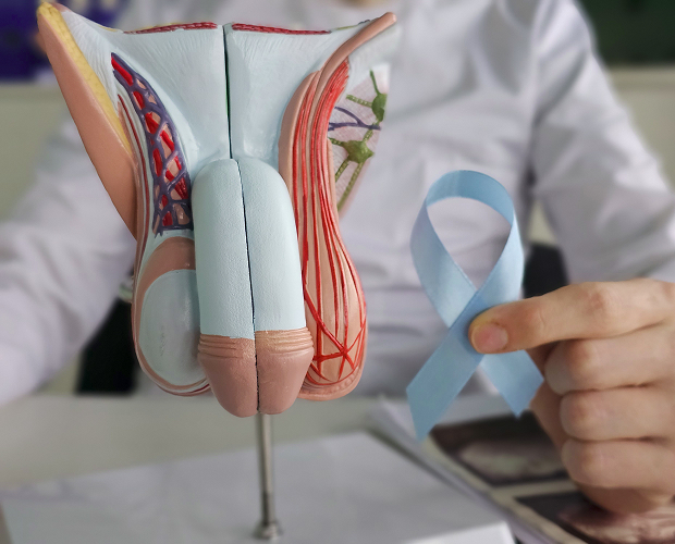
The stage is determined by the extent of the tumor and tumor marker levels; it determines the extent of treatment and prognosis. Testicular cancer stages are usually divided into three groups.
Localized testicular cancer
The testicular tumor is limited to the testicle itself. This is the most common and favorable situation, in which surgery and observation are often sufficient.
Regional spread
Tumor cells have reached the retroperitoneal lymph nodes. This process requires additional treatment after surgery, such as radiation or medication.
Distant metastases
The tumor has spread to other organs, most often the lungs. Metastases in testicular cancer are less common, but even at this stage, chemotherapy offers a high chance of cure.
General information
Treatment of seminoma
Treatment depends on the stage, but in general, this form is particularly treatable. Treatment for testicular cancer with testicular seminoma relies on three methods.
Surgical treatment
The first step is almost always surgery. Removal of the testicle along with the spermatic cord (orchiectomy) simultaneously confirms the diagnosis and removes the primary tumor.
Radiation therapy
This form is sensitive to radiation. Radiation therapy is prescribed to lymphatic drainage areas in the early stages, although it is used less frequently today than before.
Drug therapy
For advanced seminoma, chemotherapy is used. Platinum-based chemotherapy for testicular cancer is highly effective and often results in a complete cure.





Answers to frequently asked questions
What symptoms may indicate testicular cancer?
How does a seminoma differ from a non-seminoma tumor?
What tests are needed for diagnosis?
Is it possible to completely cure testicular cancer?
Yes. With timely treatment, the disease is curable in most cases, and the chances remain high even at the metastatic stage.
The outcome of testicular cancer treatment almost always depends on how quickly it is started. If you feel a lump or notice a change in your scrotum, don't delay and schedule a consultation with an oncologist.
Category of Clinical Guidelines of the Ministry of Health of the Russian Federation — https://cr.minzdrav.gov.ru/
Portal of the All-Russian National Union "Association of Oncologists of Russia" — https://oncology-association.ru
Russian Society of Clinical Oncology (RUSSCO) — https://rosoncoweb.ru
Scientific Electronic Library eLIBRARY — https://elibrary.ru
CyberLeninka — publications on oncology and oncourology — https://cyberleninka.ru
MSD Manual (Russian version) — https://www.msdmanuals.com/ru
Federal Electronic Medical Library — https://femb.ru
National Medical Encyclopedia of the Ministry of Health of the Russian Federation — https://meduniver.com
PubMed (Russian-language reviews and materials with translations for evidence-based verification) — https://pubmed.ncbi.nlm.nih.gov
Our doctors











































This award is given to clinics with the highest ratings according to user ratings, a large number of requests from this site, and in the absence of critical violations.

This award is given to clinics with the highest ratings according to user ratings. It means that the place is known, loved, and definitely worth visiting.

The ProDoctors portal collected 500 thousand reviews, compiled a rating of doctors based on them and awarded the best. We are proud that our doctors are among those awarded.
Make an appointment at a convenient time on the nearest date
Price
Other cancer treatment services

Appointment to the doctor
Reviews





















Our clinics


Application “Personal Account K+31”












Characteristics of germ cell tumors
Almost all cases are germ cell tumors, that is, neoplasms of cells that normally give rise to sperm. They are divided into two large groups, and this division determines the entire treatment strategy. The aggressive type includes non-seminomatous germ cell tumors.
What is the difference between seminoma and non-seminomatous tumors?
The difference is fundamental. Testicular seminoma grows more slowly and is sensitive to radiation. It usually occurs after the age of 30. Non-seminomatous variants—embryonic carcinoma, yolk sac tumor, choriocarcinoma, teratoma—are more aggressive and require chemotherapy. Often, a single tumor combines both components; in such cases, it is treated as a non-seminoma.