Oral Cancer (Cheek, Gum, Hard Palate, and Floor of the Mouth)
The oral mucosa is exposed daily to hot foods, sharp teeth, dentures, fillings, and accidental microtrauma. Therefore, a sore, hard spot, or stain is often perceived as a temporary irritation.

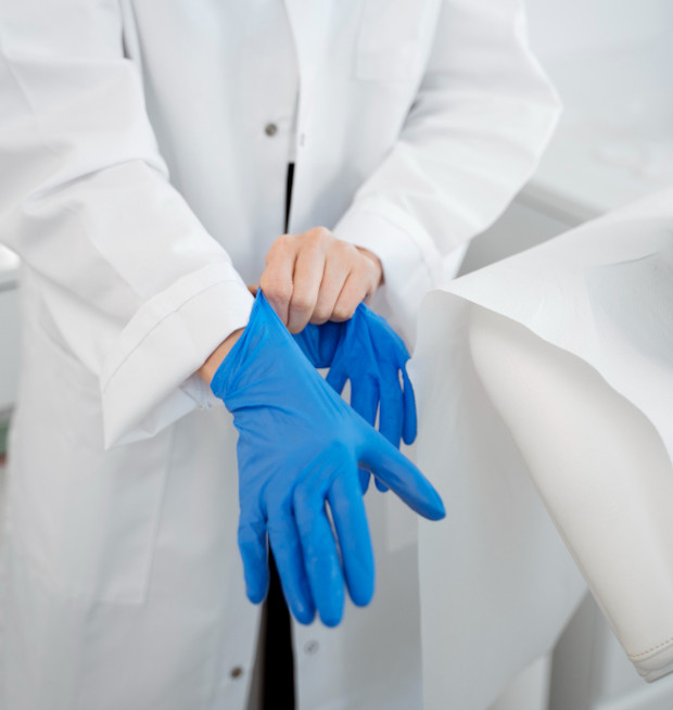
specialists

equipment

treatment
What areas of the oral cavity can be affected by tumor
Damage to different anatomical zones changes the course of the disease and requires specific surgical approaches.
Cancer of the buccal mucosa
The inner surface of the cheek is constantly in contact with teeth, dentures, fillings, and the sharp edges of crowns. Because of this, the first changes can easily be mistaken for trauma. Cancer of the buccal mucosa can begin as a dense area, a rough plaque, an ulcer with raised edges, or an area that bleeds when touched.
As the process progresses, cheek cancer affects the submucosal tissues. The lump becomes less mobile, pain develops, and chewing and opening the mouth wide becomes more difficult. When the oral tumor spreads to the masticatory muscles, limited jaw movement becomes an important diagnostic sign.
Gum cancer
In its early stages, gum cancer may appear as inflammation from a toothbrush injury, a poorly fitting denture, or a chronic root infection. Unexplained bleeding, a dense, growing area, an ulcer, an unpleasant odor, and tooth mobility near the lesion are warning signs.
Gum cancer is dangerous because it can quickly spread to the alveolar process of the jaw—the bony part where the tooth sockets are located. Therefore, if a suspicious mass is detected, the doctor evaluates the mucosa, the condition of the teeth, the bone, and the regional lymph nodes. A dental examination alone is sometimes insufficient: if the diagnosis is unclear, an oncological diagnosis is necessary.
Cancer of the hard palate
It usually begins as a small, dense lesion on the upper roof of the mouth. Initially, it may cause little pain. A person may notice a roughness in the tongue, feel discomfort when eating, or notice a discolored area on the mucous membrane.
With further growth, the lesion can destroy the palatine plate—the thin bony partition between the mouth and nasal cavity. This can cause a nasal voice, difficulty breathing through the nose, discharge, and pain in the upper jaw. These symptoms indicate a deep spread of the disease and require urgent examination.
Cancer of the floor of the mouth
The disease affects the mucous membrane under the tongue. Due to the abundant blood supply and lymph drainage of this area, oral floor cancer metastasizes to the submandibular lymph nodes very early.
|
Significant |
Benign tumor |
Malignant tumor of the mucosa |
|---|---|---|
|
Growth rate |
Slow, over years |
Fast, weeks to months |
|
Painfulness |
Usually painless |
Pain may develop as the lesion grows. |
|
Tendency to bleed |
Rarely |
Frequent (even with light touch) |
|
Change in size |
Stable or very slow growth |
Enlarges, infiltrates tissue |
|
Lymph node involvement |
Uncommon |
Common (regional metastases) |
|
Biopsy required |
As indicated |
Required for morphological verification |

Causes and risk factors
There are several factors that trigger mechanisms of malignant transformation.
Smoking and tobacco use
Tobacco smoke and smokeless tobacco contain carcinogens that cause constant chemical and thermal irritation of the epithelium, significantly increasing the risk of developing the disease.
Alcohol consumption
Ethyl alcohol acts as a strong solvent, increasing the permeability of cell membranes to other external carcinogens.
Chronic trauma to the mucosa
Constant mechanical damage to the epithelium triggers continuous cell division to heal the defect, which increases the risk of genetic errors.
Viral and Precancerous Diseases
Highly oncogenic human papillomavirus infections, as well as chronic precancerous diseases such as leukoplakia, erythroplakia, and lichen planus, play a significant role in oncogenesis.
Hereditary Factors and Associated Diseases
A general genetic predisposition plays a significant role. Chronic inflammatory processes in the oral cavity and poor hygiene create favorable conditions for the development of atypical cells.

What signs may indicate a tumor process in the mouth
The disease begins unnoticed, so it's important to know the main signs of oral cancer.
Early Signs
In the initial stages, the disease manifests itself with the appearance of ulcers, cracks, or growing white and red spots on the mucous membrane.
Symptoms of Disease Progression
As the tumor spreads into deeper tissues, the patient develops obvious symptoms of oral cancer. A constant dull or shooting pain appears, and an unpleasant putrid odor comes from the mouth.
When to see a doctor urgently
You should consult an oncologist if you notice:
- A non-healing mouth ulcer or deep crack in the mucous membrane
- A dense growth on the mucous membrane or a nodule in the soft tissue
- White or red spots that change texture
- Bleeding from the lesion without an obvious cause
- Constant pain when chewing, swallowing, or moving the tongue
- Numbness or loss of sensation in certain areas of the mucous membrane
- Densely enlarged lymph nodes in the neck or under the jaw.
To assess the spread of cancer, doctors use the TNM staging system. This staging system is very important for choosing treatment strategies.
Stage I
A tumor up to two centimeters in size is localized only in the mucosal layer, does not grow deeper into the tissue, and has not affected the lymph nodes. At this stage, early diagnosis of cancer guarantees the best treatment outcomes.
Stage II
The primary lesion increases to two to four centimeters, but the tumor is still limited to one area, has not spread to adjacent organs, and has not metastasized to the cervical lymph nodes.
Stage III
The lesion is larger than four centimeters, or the tumor is smaller, but there is one mobile metastasis up to three centimeters in a lymph node on the affected side. At this stage, patients experience obvious pain and difficulty eating.
Stage Four
This is a malignant tumor of the oral cavity that reaches large sizes and grows into the surrounding jawbones, deep neck muscles, or facial skin.
Our advantages
By choosing the oncology service at K+31 Clinic, you entrust your health to a team of experts using the most advanced medical advances.
Multidisciplinary approach
Each clinical case is discussed by a panel consisting of an oncologist, a head and neck surgeon, a medical oncologist, and a radiologist. This eliminates subjective errors and allows us to develop the most effective treatment for oral cancer.
Modern diagnostic facilities
All necessary high-precision examinations, including MRI, CT, endoscopy, and urgent histological examination of biopsies, are performed in one location at our clinic.
Personalized treatment plan
We don't use standard treatment plans; instead, we select a combination of treatment methods based on the histological subtype, stage of the disease, the patient's age, and the presence of comorbidities.
Patient support at all stages
The medical staff at K+31 Clinic provides continuous patient support.
Diagnostics
At the K31 Clinic, oral cancer diagnosis is performed using clear diagnostic algorithms and high-tech, expert-class equipment.
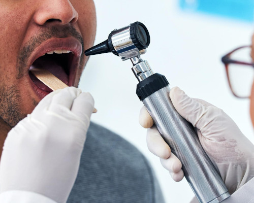
Oncologist Examination
A specialized oral oncologist performs a thorough visual examination of all parts of the oral cavity under good lighting and also performs bimanual palpation of the lesion to determine its boundaries and density.
Endoscopic Examination
We use flexible endoscopy to assess the condition of hard-to-reach areas, such as the base of the tongue, laryngopharynx, and nasopharynx.
Biopsy and Histological Examination
A mandatory step is an oral biopsy, during which the doctor removes a small sample of tumor tissue from the affected area. The collected material is sent for histological examination, where the laboratory determines the tumor's cell type.
CT, MRI, and PET-CT
Computer tomography (CT) of the facial skeleton helps to visualize the destruction of jaw bone tissue, magnetic resonance imaging (MRI) of the soft tissues of the neck provides detailed visualization of the boundaries of muscle infiltration, and positron emission tomography (PET-CT) is used to search for distant metastases throughout the body.
Laboratory Tests
Before therapy, the patient undergoes comprehensive laboratory tests. The diagnostic results are used to assess their overall health.


In our practice, we achieve the best results with patients who seek medical attention at the first sign of symptoms. Modern diagnostic methods allow us to accurately determine the extent of the disease and select a personalized treatment plan with the help of specialists from various fields.
Remember that self-diagnosis and the use of traditional treatments when cancer is suspected are unacceptable and a waste of time.
If you have any doubts or need a second expert opinion, schedule an appointment with the oncology service at K+31 Clinic.
Answers to frequently asked questions
What should be a warning sign?
Wounds that won't heal for a long time or strange lumps are a reason to see a doctor. Is the wound bleeding? Then you need to make an emergency appointment!
Is it possible to cure it completely?
In the first or second stages, the likelihood of recovery and achieving long-term remission is significantly higher.
What tests are needed to make a diagnosis?
The main methods are an in-person examination by an oncologist with palpation, an oral biopsy followed by histological examination, and imaging techniques.
Our doctors


This award is given to clinics with the highest ratings according to user ratings, a large number of requests from this site, and in the absence of critical violations.

This award is given to clinics with the highest ratings according to user ratings. It means that the place is known, loved, and definitely worth visiting.

The ProDoctors portal collected 500 thousand reviews, compiled a rating of doctors based on them and awarded the best. We are proud that our doctors are among those awarded.
Make an appointment at a convenient time on the nearest date
Price
Other cancer treatment services

Appointment to the doctor
Reviews


Our clinics

Application “Personal Account K+31”


Экстренная помощь

Скачайте приложение «Личный кабинет К+31»


Приложение «Личный кабинет К+31»


How does malignant oral mucosal disease develop?
The oncology department at K+31 Clinic pays special attention to such changes: a persistent defect, bleeding, thickening, or discoloration of the mucous membrane may look like stomatitis or a bite mark, although they sometimes indicate malignant tissue growth.
Statistics show that oral cancer is a significant cause of head and neck cancer. The success of treating this pathology depends entirely on the time of detection of the primary lesion.
The cancerous tumor grows very quickly and affects the muscles and bones of the jaw. It then metastasizes to the lymph nodes of the neck.
In the early stages, the pathological formation is painless and does not cause any discomfort. In other words, it does not cause any concern. This is why patients often seek medical attention too late.