Pancreatic cancer

Pancreatic cancer is a malignant tumor formed from the altered cells of the organ that secretes substances necessary for the digestion process and regulation of metabolism.

Best
specialists
Expert
equipment
Advanced diagnostic
treatment
Sign up
Fill out the entry form on the page and we will call you back
SUBMIT YOUR APPLICATION Позвонить
Download the application “Personal Account K+31”
Everything about your health in one click!
Subscribe to social networks K+31
To keep up to date with current offers and receive useful advice about your health.

About the disease

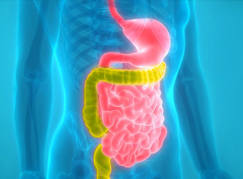
The pancreas is located in the left upper quadrant near the stomach and duodenum. It is penetrated by excretory ducts, vessels, and nerves, covered by a connective tissue capsule, and consists of three parts: the head, body, and tail. The excretory duct joins the bile duct at the head of the pancreas and opens into the duodenum.

What is the pancreas?

The main function of this organ is to produce pancreatic juice, rich in digestive enzymes (amylase, lipase, protease). These substances enter the lumen of the duodenum, where they actively break down complex protein, fat, and carbohydrate molecules into smaller compounds, ensuring normal digestion. At the same time, special cell clusters—the islets of Langerhans—produce vital hormones: insulin and glucagon. These are directly released into the blood and regulate carbohydrate metabolism, maintaining optimal glucose levels. Thus, the pancreas is an indispensable component of both the digestive and endocrine systems.

As cells divide, mutations can occur, and some of these mutations can disrupt the functions that control cell division. Then, the cells begin to divide uncontrollably, forming a tumor.

Pancreatic cancer is one of the most aggressive types of cancer. It often remains asymptomatic until the late stages of the disease, when treatment becomes very difficult. Cancer cells can spread throughout the body through the bloodstream, forming distant secondary tumors (metastases).

The five-year survival rate (the percentage of patients who survive five years after diagnosis) for early-stage pancreatic cancer is up to 15%. This is because the organ is located in a difficult-to-reach place for surgery and is closely surrounded by other organs. Surgeons must be highly experienced in performing malignant tumor removal procedures. The risk of complications after surgery is very high.

Pancreatic cancer is the sixth most common cancer and accounts for 7% of cancer deaths of all types. It most often affects older adults of both sexes. In more than half of cases, the tumor develops in the head of the pancreas. And in almost a third of cases, the entire organ is affected.

Get examined at our clinic to quickly detect diseases of the pancreas and other organs.

Schematic representation of the pancreas with the tumor marked

Prevalence of the Disease

Pancreatic cancer is one of the most pressing and complex problems in modern oncology. According to global statistics, this malignant neoplasm consistently ranks sixth in frequency of diagnosis among all oncological diseases. However, its true danger is revealed in its mortality rate – it accounts for approximately 7% of all cancer deaths, due to its extremely aggressive course and late detection.

The situation in Russia reflects global trends, demonstrating a steady increase in the number of new cases. This disease largely affects older patients, although its incidence has also been increasing in younger patients in recent years.

Regarding the localization of the pathological process, in more than half of all clinical cases, the tumor is detected in the head of the pancreas. In almost a third of patients, by the time of initial diagnosis, the disease has already reached widespread distribution, affecting the entire organ, which significantly limits radical treatment options and worsens the prognosis for each individual patient.

Experience an examination at the K+31 Clinic in Moscow to quickly detect diseases of the pancreas and other organs. We offer a full range of modern diagnostic procedures using expert-class equipment. We conduct a comprehensive analysis of all data during a consultation and provide accurate diagnoses in a short time.

Types of pancreatic cancer

MRI of the abdomen showing signs of pancreatic cancer

Based on its secretory function, the pancreas is divided into exocrine and endocrine parts. The exocrine part secretes digestive enzymes, which are released through ducts into the duodenum. Endocrine tissue secretes hormones that go directly into the blood. Depending on which of these tissues the tumor formed in, exocrine malignant tumors of the pancreas are distinguished (the most common) and endocrine.

The most common endocrine tumor is adenocarcinoma. It can be ductal (formed in the glandular cells of the pancreatic ducts - the most common form of cancer) or acinar-cell (from the cells of the acinus - a special structure of the pancreas).

In addition to adenocarcinoma, pancreatic cancer varieties include such rare types as squamous cell carcinoma, undifferentiated cancer, cystadenocarcinoma, adenosquamous carcinoma, signet ring cell carcinoma.

Visualization of pancreatic tumor in a medical examination

Benign tumors can form in the pancreas, such as intraductal papillary tumor or cystic mucinous tumor. But they can degenerate into malignant tumors, so they are often removed surgically, especially if their location interferes with the function of the pancreas. Patients with such diagnoses should be examined regularly.

Endocrine cell tumors occur in less than 5% of cases. They are called neuroendocrine tumors, as well as islet cell tumors. According to the hormone secreted by the cells responsible for the formation of the tumor, neoplasms are called: gastrinoma (a tumor of cells producing gastrin), insulinoma (insulin), glucagonoma (glucagon), somatostatinoma (somatostatin). Usually these are benign tumors of the pancreas, but there are also malignant ones.

In rare cases, malignant tumors in the pancreas are metastases from cancer in another organ - this is metastatic cancer. The primary tumor can be in the gastrointestinal tract, kidneys, lungs, mammary gland or prostate gland. Sometimes the pancreas can be affected by metastases of melanoma (skin and mucous membrane cancer), osteosarcoma (bone cancer), leiomyosarcoma (smooth muscle cancer), Merkel cell carcinoma (skin cancer).

Location of pancreatic cancer

As already mentioned, the pancreas anatomically consists of a head (adjacent to the duodenum), which passes into the body (between the duodenum and the stomach) and ends in a tail (reaches the spleen). A malignant tumor can form in any of these sections.

Cancer of the head of the pancreas

This is the most common type of pancreatic cancer by localization. Tumors in this part are smaller than in the body and tail, but due to the bile duct that passes here, even a small tumor can become a serious problem. It can compress the duct, which causes mechanical jaundice. A neoplasm in the head of the pancreas can also lead to a narrowing of the lumen (stenosis) of the duodenum. Another complication of malignant tumors of this localization is bleeding due to tumor decay.

Pancreatic body cancer

In terms of prevalence of pancreatic tumors, its body is in second place. Neoplasms here are larger than in the head. They can lead to inflammation of the vessels (thrombophlebitis), blockage of the lumen of the vessels (phlebothrombosis) and even diabetes. Advanced forms of pancreatic cancer localized in the body of this organ can cause jaundice.

Pancreatic tail cancer

This is the rarest type of pancreatic cancer in terms of location. But it is more difficult to diagnose using ultrasound.

An oncologist discusses pancreatic cancer diagnosis with a patient
Stages of pancreatic cancer

According to the international TNM system, pancreatic cancer is divided into stages depending on the characteristics of three indicators:

  • T – the degree of growth of the primary tumor: T1 – a tumor that has affected only the tissues of the pancreas up to its capsule, T2 and T3 – tumors at different stages of growth into the capsule and surrounding tissues and organs.
  • N – the number of affected lymph nodes: N0 – the lymph nodes are not affected by the tumor process, N1, N2 and N3 – cancer has affected a different (increasing) number of lymph nodes.
  • M – the presence of distant metastases: M0 – there are none, M1 – there are.

According to these indicators and their values, four stages of pancreatic cancer can be distinguished:

  • Stage 1 – T1N0M0;
  • Stage 2 – T1N1M0;
  • Stage 3 – T1N2M0, T2N0M0, T2N1M0;
  • Stage 4 – any values ​​of T or N, but necessarily M1.
Consultation at a pancreatic cancer treatment clinic in Moscow
Causes of pancreatic cancer

All cancers are caused by mutations in cells. There are many risk factors that can trigger such mutations. The most common risk factors for pancreatic cancer are:

  • Type 2 diabetes
  • Chronic pancreatitis – various inflammatory processes in the pancreas
  • Gallstone disease
  • Liver cirrhosis
  • Cysts or adenomas in the prostate gland
  • Smoking and alcohol consumption
  • Predominance of fatty and spicy foods in the diet
  • Burdened heredity
  • Excess weight, especially excess fat in the waist area
  • Age – the risk increases after 45 years, and most often the disease occurs at the age of 70 years
Anatomical image of the pancreas with the tumor area highlighted

Clinical presentation

The disease is characterized by a long asymptomatic course, making timely diagnosis difficult. Clinical manifestations become noticeable primarily when the tumor has significantly grown and spread beyond the pancreas.

Early symptoms

The initial stage of the disease often masquerades as common gastrointestinal disorders. Pain is present, but intermittent and subtle. Patients in the early stages of cancer may also experience:

  • Unexplained weight loss while maintaining a normal diet
  • Occasional discomfort in the epigastric region
  • Periodic pulling sensations in the lumbar region
  • Changes in food preferences with an aversion to fatty foods
  • Dyspeptic symptoms: flatulence, nausea, irregular bowel movements

The patient's general condition is often accompanied by unexplained weakness, apathy, and decreased performance.

Late symptoms

As the cancer progresses, the clinical picture becomes more pronounced and specific.

  1. Mechanical jaundice develops: the skin, sclera, and mucous membranes take on an intense yellowish tint, urine darkens, and stools become acholic (discolored). This process is accompanied by debilitating itching.
  2. Pain intensifies, becoming constant and excruciating, especially if the tumor invades the nerve plexuses.
  3. Cancer intoxication develops, leading to severe cachexia (wasting) and persistent fever.
  4. Metastasis to the peritoneum can lead to ascites—fluid accumulation in the abdominal cavity.
  5. The risk of thrombosis increases. In particular, there is a high risk of developing migratory thrombophlebitis (Trousseau's sign). A sign of this condition is the appearance of painful redness and hardening along the veins in various parts of the body, which may disappear on their own and then reappear.
  6. If the endocrine part of the organ is affected, secondary diabetes mellitus may manifest. This is characterized by sudden intense thirst, dry mouth, and increased urine output.

Any of these symptoms is a serious reason to immediately consult an oncologist.

Diagnosis of pancreatic cancer

The difficulty of early detection is due to the long asymptomatic development of the pathology. By the time pronounced clinical symptoms appear, the tumor process often reaches a widespread stage with metastasis. Modern pancreatic cancer diagnostics requires a comprehensive approach using laboratory and instrumental techniques.

Laboratory diagnostics

Laboratory diagnostics provide valuable information about the functional state of the pancreas and systemic changes in the body characteristic of oncology. Blood and biological fluid tests can identify specific markers indicating the presence of a pathological process. The following are used to diagnose cancer:

  • Blood tests for pancreatic enzymes, bilirubin metabolism parameters, and liver function tests
  • Tumor marker testing, particularly CA 19-9, although its specificity requires further confirmation.
  • Coagulation profile evaluation to identify associated disorders
  • Molecular genetic testing to determine targeted therapy tactics
  • Liquid biopsy – detection of circulating tumor cells and DNA in the blood

Instrumental diagnostics

Visualization of pancreatic structures and surrounding tissues is a key step in confirming the diagnosis. Modern instrumental methods allow us to accurately determine the location, size, and nature of the tumor, as well as assess the extent of the process.

The patient undergoes:

  • Abdominal ultrasound is a basic screening diagnostic method for primary complaints.
  • Endoscopic ultrasound (endosonography) provides detailed visualization of the pancreas and allows for fine-needle biopsy.
  • Contrast-enhanced computed tomography determines the size, location, and relationship of the tumor to surrounding structures.
  • Magnetic resonance imaging with cholangiopancreatography protocols (MRCP) provides detailed information about the condition of the pancreatic and bile ducts.
  • ERCP combines endoscopic and radiological methods, allowing for tissue collection and stent placement.
  • PET-CT detects distant metastases and Assesses the extent of the process.
  • Angiography – determines the characteristics of the tumor's blood supply and its relationship to the main vessels.

For a definitive diagnosis, histological confirmation of the malignancy of the tumor is required by biopsy, performed under ultrasound or CT guidance.

General information

Pancreatic cancer tumor markers

When oncological processes occur in the body, the level of some substances in the blood increases. Such substances are called tumor markers. For pancreatic cancer, these are: CA 19-9, CEA, CEA. But tumor marker tests do not provide 100% certainty. Since their level can fluctuate for other reasons. However, for monitoring the treatment process, they can be a very informative indicator of the condition and effectiveness of the chosen therapy method. For example, an increase in the level of tumor markers during cancer treatment may indicate that the therapy is not giving results. And an increase after treatment is a sign of a relapse.

Modern equipment for diagnosing pancreatic oncology

Pancreatic cancer metastases

Approximately half of patients with pancreatic cancer have metastases, which corresponds to stage 4 of the disease. These tumor foci are located in different organs of the abdominal cavity. They can be in the liver or lungs and even in the bones. Unfortunately, there is no effective treatment for pancreatic cancer with metastases. In such cases, palliative care is provided, i.e. treatment is aimed at reducing pain, containing tumor processes and prolonging the patient's life.

Pancreatic carcinoma under a microscope

Treatment of pancreatic cancer

A panel of doctors selects therapy based on the stage, type of pancreatic cancer and other features of the disease. Treatment is complicated by the fact that cancer is most often detected at late stages, when the oncological process has spread widely.

The main methods of treating pancreatic cancer:

  • Surgical. In a small percentage of cases, when there are no metastases, neighboring organs are not affected and the patient's health allows, this method is possible. The type of surgery depends on the location of the malignant tumor. If the tumor is in the head of the pancreas, then the Whipple procedure (pancreatoduodenectomy) is most often performed. Malignant lesions of other parts of the gland may require distal pancreatectomy. If it is necessary to remove the entire organ (as well as the gallbladder, parts of the stomach and intestines, and the spleen), then a total pancreatectomy is performed
  • Nano-knife is a new technology, which is also called irreversible electroporation. Electrodes are placed into the tumor under ultrasound or CT control, and its cells are destroyed under the influence of electrical impulses. This method is used when conventional surgical removal is impossible for various reasons. And it refers to palliative treatment methods. The risks of such an intervention are lower, but it does not cure pancreatic cancer, but can only prolong the patient's life
  • Chemotherapy. This is not the most effective method of treating pancreatic cancer. Chemotherapy drugs affect the tumor, completely destroying or reducing its size. These can be intravenous infusions, tablets, or both. Most often, chemotherapy is used in combination with radiation therapy and usually as a palliative treatment
  • Radiation therapy. The method is widely used to treat cancerous growths, including pancreatic tumors. Rays are used before or after surgery, most often together with chemotherapy. Before surgery, the goal of radiation therapy is to reduce the size of the tumor. After surgery, it is to kill the cancer cells that remain after the removal of the malignant tumor. Also, a combination of chemotherapy and radiation therapy is used when surgery is not possible and the patient needs to be relieved of pain

The following are also used to treat pancreatic cancer:

  • Targeted therapy. These types of drugs work only on malignant tumor cells, without damaging normal cells
  • Immunotherapy. This is a cancer treatment method that is designed to stimulate the body's immune system to better recognize cancer and fight it
  • Proton therapy. Effective local action on a malignant tumor destroys cancer cells. This method is not yet used in Russia

The use of certain methods of treating pancreatic cancer depends on the type, stage, and other features of the disease. The therapy strategy is selected by a council of doctors.

A lab technician performs tests to detect a pancreatic tumor.

Pain relief during treatment

Pain is one of the most severe and debilitating manifestations of the disease, significantly reducing the patient's quality of life. Intense pain occurs due to tumor growth into the richly innervated capsule of the gland and surrounding nerve plexuses, as well as due to compression of adjacent organs and the development of inflammation.

A stepwise approach is used to relieve pain:

  • Nonsteroidal anti-inflammatory drugs are prescribed initially.
  • Weak opioid analgesics are effective for moderate pain.
  • Strong, long-acting opioid drugs are indicated for severe pain.

If drug therapy is ineffective, innovative interventional techniques are used to relieve symptoms:

  • CT-guided percutaneous celiac plexus block
  • Endoscopic ultrasound neurolysis
  • Radiofrequency ablation of nerve ganglia

In parallel with analgesic therapy, Symptomatic treatment, including enzymatic drugs to reduce the functional load on the organ and anti-inflammatory therapy. An individually tailored pain management regimen can significantly improve the patient's condition and create conditions for primary antitumor treatment.

Also used for the treatment of pancreatic cancer are:

  • Targeted therapy. These drugs act only on malignant tumor cells, without damaging normal cells.
  • Immunotherapy. This cancer treatment method is designed to stimulate the body's immune system to better recognize and fight cancer.
  • Proton therapy. Effective local action on the malignant tumor destroys cancer cells. This method is not yet used in Russia.

The use of specific treatment methods for pancreatic cancer depends on the type, stage, and other characteristics of the disease. A panel of doctors determines the treatment strategy.

A patient undergoing treatment for pancreatic cancer at a medical center

Possible complications

The progression of pancreatic cancer leads to the development of serious complications that significantly worsen the patient's condition and force doctors to change treatment strategies. These pathological conditions arise both as a result of local tumor spread within the organ and as a result of systemic effects on the body.

  1. Mechanical jaundice. Develops when the bile ducts are obstructed by a tumor mass localized in the head of the pancreas. Impaired bile flow leads to the accumulation of bilirubin in the blood, manifesting as a characteristic triad. This includes intense yellowing of the skin and sclera, acholic stool, and dark urine. Prolonged jaundice is accompanied by itching and leads to impaired liver detoxification, posing a risk of liver failure.
  2. Pancreatogenic cachexia. Characterized by progressive depletion of the body due to a combination of factors. Impaired exocrine function of the organ leads to a deficiency of digestive enzymes and malabsorption syndrome. Simultaneously, the tumor provokes a systemic inflammatory response with increased energy expenditure, which, combined with digestive dysfunction, creates a specific metabolic state resistant to standard nutritional support.
  3. Intestinal obstruction. Occurs with invasive tumor growth into the duodenum or compression of intestinal loops by a space-occupying lesion. It presents with a complex of characteristic symptoms. These include cramping abdominal pain, persistent vomiting, and prolonged stool retention with no flatulence. This condition requires urgent surgical intervention to restore intestinal movement.
  4. Secondary diabetes mellitus. Destruction of insulin-producing cells of the pancreas by the tumor process leads to the development of a specific diabetes status. The disease is characterized by a labile course with a tendency toward hypoglycemic states, which is due to concomitant hormone deficiency. Its characteristic feature is resistance to standard hypoglycemic therapy.
  5. Profuse bleeding. This may occur in the late stages due to erosion, that is, damage to the integrity of large vessels in the retroperitoneal space, or due to tumor penetration into the gastrointestinal tract. It is characterized by massive blood loss with the development of hemorrhagic shock, which manifests as severe weakness, dizziness, tachycardia, and a drop in blood pressure. This complication is immediately life-threatening and requires emergency surgical intervention.

Each of these complications requires prompt diagnosis and active medical care in a specialized hospital with comprehensive treatment and intensive care capabilities.

Surgical intervention for pancreatic oncology

Prognosis

Prognostic indicators for this pathology remain quite low, due to late diagnosis and the aggressive biological behavior of the tumor. Five-year survival rates across all stages generally do not exceed 9-15%, but vary significantly depending on the stage of disease detection.

In localized forms, when radical surgery is possible, this rate reaches 30-35%. At advanced stages with distant metastases, the median survival is approximately 6-11 months, even with chemotherapy.

Prognostic outlook and survival time are influenced by a number of factors:

  • Disease stage and the possibility of radical surgery
  • Tumor differentiation level
  • Patient age and presence of comorbidities
  • General condition according to the ECOG/WHO scale
  • Response to drug therapy
  • Molecular genetic characteristics of the tumor

Timely surgical intervention, including pancreaticoduodenectomy, remains a key factor in significantly improving long-term outcomes. Modern treatment protocols can improve the prognosis and increase median survival by 4-6 months in advanced forms of the disease.

Risk of recurrence

The risk of cancer recurrence remains significant even after successful completion of primary treatment. More than 70% of patients who have undergone radical surgery experience locoregional recurrence or distant metastases within the first two years.

Key patterns of recurrent cancer:

  1. The most common sites are the body of the pancreas, liver, and peritoneum.
  2. The peak incidence occurs in the first 18-24 months of follow-up.
  3. The risk correlates with the initial stage and the extent of surgery. The later the tumor is detected, the higher the risk.
  4. The risk of recurrence is higher in the presence of mutations in the BRCA1/2 genes.

The standard follow-up protocol after successful treatment includes mandatory regular examinations:

  • The first 2 years – monitoring every 3-4 months
  • From years 3 to 5 – monitoring every 6 months
  • After 5 years – annual monitoring.

The monitoring program includes abdominal CT, CA 19-9 tumor marker testing, and ultrasound. If an isolated recurrence is detected, the possibility of repeat surgery or radiofrequency ablation is considered. In the case of disseminated disease, systemic drug therapy is indicated based on the tumor's molecular profile.

Prevention of pancreatic cancer

Prevention measures for this oncological disease that a person can influence include: a healthy lifestyle and giving up bad habits, maintaining a normal weight, observing safety rules in hazardous production to limit contact with harmful substances.

The oncology department of our clinic "K+31" is equipped with the latest equipment for the diagnosis and treatment of pancreatic cancer. Our doctors use modern proven methods, study the latest research and have extensive experience in treating different types of cancer. To visit an oncologist, call us by phone or leave a request on the website, and we will call you back as soon as possible.

A patient undergoing treatment for pancreatic cancer at a medical center

Post-treatment rehabilitation

Comprehensive recovery after aggressive anticancer treatment is a multi-stage process requiring an integrative approach. A medical rehabilitation program is developed individually, taking into account the extent of the intervention, the patient's age, and the patient's functional state.

  1. Post-Surgical Recovery. Post-operative rehabilitation includes early patient activation to prevent thromboembolic complications and pneumonia. A special kinesitherapy program is aimed at restoring abdominal muscle strength and preventing post-operative hernias. Breathing exercises and postural drainage help improve pulmonary ventilation. Gentle physiotherapy procedures help relieve pain and normalize gastrointestinal motility.
  2. Nutrition support and diet therapy. A specialized diet includes frequent meals with increased levels of easily digestible protein and limited fat. Synthetic nutritional mixtures for enteral nutrition are prescribed if necessary. Lifelong pancreatic enzyme replacement therapy is a mandatory component of rehabilitation measures, the dosage of which is individually determined based on a coprological examination and nutritional status assessment.
  3. Immunity strengthening. Immunomodulatory support after pancreatic cancer is aimed at restoring adaptive immunity and preventing the development of infections. It includes the use of prebiotics, vitamin and mineral complexes, and specialized metabolites.
  4. Psychiatric care. Psychotherapeutic support is aimed at overcoming anxiety and depressive disorders associated with the disease. Cognitive behavioral therapy helps adapt to a new quality of life, while relaxation techniques reduce stress. If necessary, mild antidepressants with minimal side effects are prescribed. Self-help groups and social integration contribute to the improvement of psycho-emotional well-being.

The rehabilitation program is implemented under the constant supervision of a multidisciplinary team of doctors, including an oncologist, nutritionist, physiotherapist, and psychologist. Regular assessment of the effectiveness of rehabilitation measures allows for timely adjustments to the strategy and optimal results.

FAQ

Which modern approaches are the most promising?

Modern tactics involve a comprehensive approach using surgical techniques, chemotherapy, and radiation therapy. Today, extended surgeries are performed using robotic systems, targeted drugs, and immunotherapy. The choice of method depends on the stage of the disease, tumor location, and the patient's overall condition.

Which diagnostic methods provide the greatest accuracy?

Combined imaging techniques are considered the most informative: multislice CT with contrast, MRI with cholangiopancreatography, and endosonography. Endosonography allows for image-guided fine-needle biopsy. PET-CT is also used to assess the extent of the disease.

How effective is surgical treatment?

Radical surgery (pancreaticoduodenectomy) remains the only method capable of achieving a complete cure. At the K+31 Clinic, such procedures are performed using minimally invasive techniques, which reduces the risk of complications.

Where should I go for pancreatic cancer treatment in Moscow?

If you have been diagnosed with or suspected of having pancreatic cancer, contact the oncology department at the K+31 Clinic in Moscow. Here, you can receive a comprehensive diagnosis, get a second opinion from leading doctors, and determine the most appropriate treatment plan for your specific case.

Our doctors

Merkulov Igor Alexandrovich
Experience 34 years
Make an appointment
Merkulov
Igor Alexandrovich
Deputy Chief Physician for Oncology, Oncologist
Ershova Ksenia Igorevna
Experience 23 years
Make an appointment
Ershova
Ksenia Igorevna
Head of department, oncologist
Abashin Sergey Yuryevich
Experience 43 years
Make an appointment
Abashin
Sergey Yuryevich
Head of oncology projects, oncologist
Kalakutskaya Natalia Lvovna
Experience 28 years
Make an appointment
Kalakutskaya
Natalia Lvovna
Doctor-oncologist
Malygin Sergey Evgenyevich
Experience 31 year
Make an appointment
Malygin
Sergey Evgenyevich
Oncologist-mammologist, surgeon
Pshikhachev Ahmed Mukhamedovich
Experience 23 years
Make an appointment
Pshikhachev
Ahmed Mukhamedovich
Urologist, Oncologist
Shevchuk Alexei Sergeyevich
Experience 26 years
Make an appointment
Shevchuk
Alexei Sergeyevich
Oncogynecology consultant, obstetrician-gynecologist
Chichkanova Tatyana Vladimirovna
Experience 25 years
Make an appointment
Chichkanova
Tatyana Vladimirovna
Oncologist-mammologist, radiologist
Ovsiy Oksana Gennadievna
Experience 14 years
Make an appointment
Ovsiy
Oksana Gennadievna
Doctor-oncologist
Katz Ksenia Vladimirovna
Experience 12 years
Make an appointment
Katz
Ksenia Vladimirovna
Dermatovenerologist, oncologist
Korshikova Kamila Mukhtorovna
Experience 5 years
Make an appointment
Korshikova
Kamila Mukhtorovna
Radiotherapist
Ter-Arutyunyants Svetlana Andreevna
Experience 27 years
Make an appointment
Ter-Arutyunyants
Svetlana Andreevna
Radiotherapist, oncologist
Udin Oleg Ivanovich
Experience 32 years
Make an appointment
Udin
Oleg Ivanovich
Deputy chief physician for surgery, surgeon
Kogonia Lali Mikhailovna
Experience 52 years
Make an appointment
Kogonia
Lali Mikhailovna
Chemotherapist
Volkova Daria Mikhailovna
Experience 17 years
Make an appointment
Volkova
Daria Mikhailovna
Head of the Department of Radiation Therapy, Radiotherapist
Gomov
Mikhail Alexandrovich
Consultant in oncogynecology, obstetrician-gynecologist
Menkes (Ryabova) Yulia Alexandrovna
Experience 5 years
Make an appointment
Menkes (Ryabova)
Yulia Alexandrovna
Oncologist-chemotherapist
Grishin Igor Igorevich
Experience 33 years
Make an appointment
Grishin
Igor Igorevich
Obstetrician-gynecologist
Achba Maya Otarovna
Experience 18 years
Make an appointment
Achba
Maya Otarovna
Radiologist, ultrasound diagnostician, oncologist-mammologist
Lukyanenko Vladimir Alexandrovich
Experience 18 years
Make an appointment
Lukyanenko
Vladimir Alexandrovich
Oncologist-mammologist
Volenko Ivan Alexandrovich
Experience 15 years
Make an appointment
Volenko
Ivan Alexandrovich
Surgeon-oncologist-mammologist, plastic surgeon
Khailova Maria Sergeevna
Experience 5 years
Make an appointment
Khailova
Maria Sergeevna
Oncologist
Peshkova Marina Sergeevna
Experience 3 years
Make an appointment
Peshkova
Marina Sergeevna
Oncologist
Yakovleva Yana Sergeevna
Experience 8 years
Make an appointment
Yakovleva
Yana Sergeevna
Radiotherapist
Ivanova Olga Vladimirovna
Experience 26 years
Make an appointment
Ivanova
Olga Vladimirovna
Radiotherapist
Magdiev Arslan Khulatdaevich
Experience 14 years
Make an appointment
Magdiev
Arslan Khulatdaevich
Surgeon, oncologist, phlebologist
Morozova Albina Soslanovna
Experience 18 years
Make an appointment
Morozova
Albina Soslanovna
Acting head of the department of antitumor drug therapy, oncologist
Iluridze Georgy Davidovich
Experience 9 years
Make an appointment
Iluridze
Georgy Davidovich
Oncologist, traumatologist-orthopedist, surgeon
Alferov Anton Sergeevich
Experience 16 years
Make an appointment
Alferov
Anton Sergeevich
Leading urologist, andrologist
Gabaraev Alan Petrovich
Experience 13 years
Make an appointment
Gabaraev
Alan Petrovich
Urologist, andrologist
Balkarov Beslan Khasenovich
Experience 17 years
Make an appointment
Balkarov
Beslan Khasenovich
Surgeon, oncologist, acting head of the surgical department
Nemenov Alexander Alexandrovich
Experience 7 years
Make an appointment
Nemenov
Alexander Alexandrovich
Urologist, oncologist (oncourologist)
Melkonyan Lia Eduardovna
Experience 13 years
Make an appointment
Melkonyan
Lia Eduardovna
Oncologist, oncologist-mammologist, surgeon, radiologist, ultrasound diagnostician
Sokorutov Vasily Ivanovich
Experience 22 years
Make an appointment
Sokorutov
Vasily Ivanovich
Oncologist
Mashkey Maria Igorevna
Experience 2 years
Make an appointment
Mashkey
Maria Igorevna
Oncologist
Dubinina Yulia Nikolaevna
Experience 13 years
Make an appointment
Dubinina
Yulia Nikolaevna
Oncologist, hematologist
Ivanov Alexey Mikhailovich
Experience 22 years
Make an appointment
Ivanov
Alexey Mikhailovich
Oncologist
All specialists
2GIS Award
2GIS Award

This award is given to clinics with the highest ratings according to user ratings, a large number of requests from this site, and in the absence of critical violations.

«Good place» according to Yandex

This award is given to clinics with the highest ratings according to user ratings. It means that the place is known, loved, and definitely worth visiting.

Our doctors are laureates of the ProDoctors Award

The ProDoctors portal collected 500 thousand reviews, compiled a rating of doctors based on them and awarded the best. We are proud that our doctors are among those awarded.

Make an appointment at a convenient time on the nearest date

Price

Reception
Price
Primary oncologist appointment
from 6 450 ₽
Repeated appointment with an oncologist
from 5 700 ₽

Appointment to the doctor

Fill out the form, our managers will contact you within 15 minutes
Почему К+31?
К + 31 — full-cycle multidisciplinary medical centers, including the possibility of providing medical services of European quality level.
К + 31 — are leading doctors and diagnostics using high-tech equipment from world manufacturers (Karl Storz, Olympus, Siemens, Toshiba, Bausch&Lomb, Technolas, Zeiss, Topcon).
К + 31 — is ethical. The staff of K+31 clinics maintain open relationships with patients and partners. An individual approach to each patient is the basis of our service standards.
К + 31 — is modernity. On call 24/7: call center operators will answer your questions at any time and book you an appointment with doctors. Contact us by phone, through the feedback form on the website and Max.

Our clinics

K+31 on Lobachevskogo

st. Lobachevskogo, 42/4

+7 499 999-31-31

Subway
1
11
Prospect Vernadsky Station
By a car
Lobachevsky, we pass the first barrier (security post of the City Clinical Hospital No. 31), turn right at the second barrier (security post K+31)
Parking pass
Opening hours
Mon-Fri: 08:00 – 21:00
Weekend: 09:00 – 19:00
K+31 West

Orshanskaya, 16/2; Ak. Pavlova, 22

74999993131

Subway
3
Molodezhnaya
By a car
Moving along Orshanskaya street, we turn to the barrier with the guard post K+31. You do not need to order a pass, they will open it for you
Opening hours
Mon-Fri: 08:00 – 21:00
Sat-Sun: 09:00 – 18:00
Didn't find the service you were looking for?

Экстренная помощь