
Neuroendocrine tumors (NETs)
This group of tumors is rare: neuroendocrine tumors grow slowly, and initial symptoms may resemble gastritis or fatigue. Only an oncologist at K+31 can understand their nature.

specialists

equipment

treatment






What are neuroendocrine tumors
These are neoplasms of cells of the neuroendocrine system: they are found in many organs, are part of the endocrine system, and produce hormones. When a cell divides uncontrollably, a neuroendocrine tumor occurs.
Characteristics of neuroendocrine neoplasms
The group is distinguished by the cells' ability to produce hormones; neuroendocrine neoplasms are classified by their degree of malignancy (Ki-67 index), which determines the prognosis.
Where can NETs occur?
Progenitor cells are present almost everywhere. Therefore, the location varies—the digestive system and pancreas are most commonly affected. Typical areas where NETs are detected are:
- Stomach, intestines, appendix, rectum
- Pancreas
- Lungs and bronchi
- Adrenal glands and paraganglia
- Less commonly—the kidneys, pelvis, and thymus gland
Complaints depend on the location. The scope of the examination—the same diagnosis may be treated differently in two patients.
Functional and nonfunctional tumors
The key distinction is based on hormonal activity. Functional, or hormonally active, tumors produce distinct syndromes: for example, carcinoid syndrome in a carcinoid tumor (the term is carcinoid). Nonfunctional tumors grow quietly and are discovered incidentally.
Symptoms of neuroendocrine tumors

The presentation depends on whether the nodule secretes hormones, its location, and its size; the symptoms of a neuroendocrine tumor can be conveniently divided into groups.
General signs of the disease
Most often, the symptoms are nonspecific—weakness, weight loss, abdominal pain; these symptoms of a neuroendocrine tumor are easily attributed to fatigue.
Hormonally active manifestations
If the tumor secretes hormones, the presentation is more complex. Carcinoid syndrome with a carcinoid tumor includes hot flashes, diarrhea, and palpitations. If the pancreas is affected, sugar spikes and stomach ulcers are present. It's all related to hormones.
Symptoms depending on location
Location dictates the symptoms: in the lungs, a cough; gastrointestinal tumors cause pain and bloating; and distant metastases of NETs are found in the liver.
Diagnosis of neuroendocrine tumors
The examination goes from simple to complex: the diagnosis of neuroendocrine tumors combines tests, visualization, and tissue examination. The patient's path is as follows:
- Oncologist consultation, collection of complaints and examination
- Blood and urine tests for specific markers
- Ultrasound, then CT or MRI with contrast
- PET-CT with somatostatin analogs as indicated
- Biopsy with morphological and immunohistochemical examination
- Analysis at a consultation and choice of tactics
Each step clarifies the previous one: without tissue, the diagnosis is not confirmed, without visualization, the tumor process cannot be assessed.
Oncologist consultation "K+31"
The doctor asks detailed questions about the patient's complaints and their progression; the NET oncologist reviews the patient's entire medical history and decides what to test next.
Laboratory research
Chromogranin A is measured, and in carcinoid syndrome, 5-hydroxyindoleacetic acid is measured; laboratory diagnostics provide guidance.
Instrumental diagnostics
They start with an ultrasound, then a CT scan and MRI. PET-CT has a special place, where the drug binds to somatostatin receptors and locates lesions.
Biopsy and morphological examination
The diagnosis is confirmed by a tumor biopsy: immunohistochemistry determines the cell type, and the Ki-67 index determines the degree of malignancy. Histology determines treatment. The methods are conveniently compared in the table.
| Method | What it shows | Advantages | Limitations | When it is prescribed |
|---|---|---|---|---|
| Ultrasound | Organ masses | Available, radiation-free | Less visible in small lesions | First stage, if complaints arise |
| CT with contrast | Dimensions and prevalence | Detailed and fast | Radiation exposure | Staging, surgical plan |
| MRI | Liver, soft tissue, invasion | Accurate, radiation-free | Longer time | For liver damage |
| PET-CT with somatostatin | Active lesions in the body | Finds hidden lesions | More expensive, not available everywhere | If spread is suspected |
| Biopsy with IHC | Cell type and grade of malignancy | Confirms the diagnosis | Invasive | Before choosing treatment |
No single method works alone. The doctors at our center pay attention to a common situation.
"In our practice, we often see that neuroendocrine tumors remain undetected for a long time due to nonspecific symptoms. Therefore, comprehensive diagnostics and collaboration between specialists in different fields are so important."
General information
Modern treatment methods for NETs
There is no single treatment: the program depends on the location, size, malignancy, and metastases, so treatment for neuroendocrine tumors is almost always a combination.
Surgical treatment
For limited disease, surgery is the primary method; surgical treatment varies, from endoscopic removal of the node to pancreatic or liver surgery.
Drug therapy
Somatostatin analogs inhibit growth and relieve hormonal symptoms; for rapidly progressing forms, chemotherapy is used—this makes treatment of neuroendocrine tumors systemic.
Targeted therapy
This approach is highly targeted: targeted therapy uses targeted drugs that block tumor division signals and vascular growth in advanced forms.
Radionuclide therapy
This method combines diagnosis and treatment: radionuclide therapy carries a therapeutic isotope that locates somatostatin receptors on tumor cells and irradiates them in a targeted manner.
Radiation therapy
External beam radiation is used less frequently for bone metastases, inoperable lesions, and pain relief; this combination makes comprehensive tumor treatment flexible.




Answers to frequently asked questions
Below are frequently asked questions from patients.
What is a neuroendocrine tumor?
What symptoms may indicate NET?
How is the diagnosis confirmed?
Can neuroendocrine tumors be treated effectively?
These growths require attention, but modern medicine can bring them under control. The earlier you begin screening, the greater your chances—don't delay your visit to the oncologist.
Clinical Guidelines of the Ministry of Health of the Russian Federation — https://cr.minzdrav.gov.ru/
Category of Clinical Guidelines of the Ministry of Health of the Russian Federation — https://cr.minzdrav.gov.ru/schema
Scientific Electronic Library eLIBRARY — https://www.elibrary.ru
CyberLeninka (publications on neuroendocrine tumors) — https://cyberleninka.ru
Russian Society of Clinical Oncology (RUSSCO) — https://rosoncoweb.ru
National Medical Encyclopedia and specialized reviews on the RMJ portal — https://www.rmj.ru
MSD Manual (Russian version) — https://www.msdmanuals.com/ru
Disease Directory on the portal "Physician's Consultant" — https://www.rosmedlib.ru
Federal Electronic Medical Library — https://femb.ru
Oncology of Russia Portal — https://oncor.ru
Russian Journal of Gastroenterology, Hepatology, and Proctology — https://www.gastro-j.ru
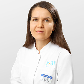
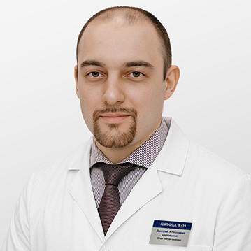
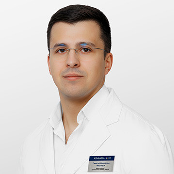
Our doctors










































This award is given to clinics with the highest ratings according to user ratings, a large number of requests from this site, and in the absence of critical violations.

This award is given to clinics with the highest ratings according to user ratings. It means that the place is known, loved, and definitely worth visiting.

The ProDoctors portal collected 500 thousand reviews, compiled a rating of doctors based on them and awarded the best. We are proud that our doctors are among those awarded.
Make an appointment at a convenient time on the nearest date
Price
Other cancer treatment services

Appointment to the doctor
Reviews





















Our clinics


Application “Personal Account K+31”












Causes and risk factors for the development of NETs
The exact cause is often unknown, but certain conditions are known to increase the risk.
Hereditary syndromes
Some of these neoplasms are associated with hereditary syndromes (MEN 1 and 2, von Hippel-Lindau disease); it is important to tell your doctor about these in your relatives.
Genetic features
Even outside of these syndromes, genetic defects are found in cells, which is why cancer diagnostics include molecular genetic analysis.
Other risk factors
Other factors have been less studied and rarely act alone: chronic inflammation, gastric diseases with low acidity, age, and smoking.