Inflammation of the Salivary Glands (Sialoadenitis)
Sialoadenitis is an inflammatory process occurring in the salivary gland tissue. The parotid or submandibular structures are most commonly affected. The disease manifests as painful swelling in the cheek or under the jaw, dry mouth, difficulty swallowing, and is often accompanied by fever. Experts estimate that inflammatory diseases of the salivary glands account for 5% to 10% of all maxillofacial pathologies in adults, with the risk increasing after age 50-60.
Treatment strategies for sialoadenitis vary. Conservative antibiotic therapy and physiotherapy are appropriate for severe symptoms, while surgical interventions are recommended if complications develop. Sialoadenitis treatment is provided at the K+31 multidisciplinary clinic in Moscow. We utilize comprehensive diagnostic and therapeutic protocols to quickly relieve symptoms and prevent relapse.

specialists

equipment

treatment


Causes and factors provoking the disease
The development of the disease is always associated with the influence of one or more pathogenic factors, which can be divided into direct causes and conditions that create a favorable environment for the disease.
Direct causes of inflammation include:
- Bacterial infections. The disease is most often caused by microorganisms normally present in the oral cavity – staphylococci and streptococci. When local immunity is weakened, they penetrate the gland ducts, causing an inflammatory reaction.
- Viral agents. A classic example is the mumps virus, which selectively affects the parotid glands. Cytomegalovirus, coxsackievirus, and influenza viruses can also cause sialadenitis.
- Obstruction of the saliva ducts. The glands can become inflamed due to obstruction of the salivary flow due to mechanical obstruction. The most common cause of obstruction is sialolithiasis, or the formation of calcium stones in the ducts. The submandibular glands are primarily affected. The ducts can also be blocked by thick mucus or compressed from the outside by edema or tumor.
Risk factors cannot be considered a direct cause, but they significantly increase the likelihood of developing sialadenitis. The key one is xerostomia, or excessive dry mouth. It occurs as a result of:
- Chronic diseases: Sjogren's syndrome, diabetes, renal failure
- Medications: diuretics, antihistamines, antihypertensives
- Dehydration
- The postoperative period, especially after abdominal surgery
- Neglect of oral hygiene
Notably, in children, the disease is more often associated with viral infections, while in adults, bacterial forms predominate, caused by duct obstruction or decreased salivary flow due to chronic pathologies.

Development of the pathological process
The trigger is most often a decrease in the rate of salivary flow and obstruction of salivary drainage. When saliva production slows and its flow is blocked, ideal conditions are created for ascending infection. Bacteria and viruses from the oral cavity easily penetrate the duct and move against the flow of saliva deeper into the gland.
Then, the microorganisms begin to actively multiply. This causes a local inflammatory reaction. The tissues of the gland and duct swell, further aggravating the obstruction of salivary drainage. Leukocytes rush to the site of infection, causing hyperemia and painful infiltration. Bacterial waste products and dead cells form a purulent exudate, which can accumulate in the lobules of the gland, leading to the formation of an abscess. Beginning as serous, the inflammation, if left untreated, quickly progresses to a purulent form, which is accompanied by increased pain, intoxication, and the risk of spreading infection to surrounding tissues.

Types of pathology
To select the most effective treatment strategy, all pathological variants are classified according to several criteria. This classification allows the physician to determine the nature of the disease, its stage, and the extent of its progression.
Based on the course of the disease, two main forms are distinguished:
- Acute. Characterized by a sudden onset and pronounced symptoms. Most often, it is infectious in nature and, with adequate treatment, results in full restoration of glandular function.
- Chronic. Occurs with alternating periods of remission and exacerbations. During the remission phase, symptoms may be absent or manifest as minor discomfort. This form is often associated with autoimmune processes, persistent decreased salivation, or anatomical features of the ducts.
Based on the etiology, sialadenitis is divided into:
- Viral
- Bacterial
- Autoimmune
- Obstructive
Depending on the location of the inflammatory process, the following are diagnosed:
- Mumps – damage to the parotid glands
- Submandibulitis – inflammation of the submandibular glands
- Sublingivitis – a rare pathology affecting the sublingual glands

Clinical signs
The clinical picture directly depends on the form of the disease. In acute sialadenitis, symptoms develop rapidly and are intense. The leading symptom is sharp pain in the affected gland, which intensifies significantly with eating and swallowing. The gland itself increases in size, which is visually manifested as a pronounced, painful swelling, often accompanied by redness of the overlying skin. Another characteristic symptom of an active pathological process is the discharge of pus from the opening of the duct when pressure is applied to the gland. Signs of intoxication include fever, weakness, and headache.
The chronic form presents differently. Pain is mild. Patients often describe it as a feeling of discomfort or heaviness. The main symptom is recurrent swelling of the gland, which appears or intensifies during meals. Between flare-ups, the gland's function gradually declines, leading to persistent dry mouth. Long-term chronic inflammation can lead to irreversible changes—fibrosis and atrophy of the glandular tissue, when the gland shrinks in size and virtually ceases secreting.
Diagnostic methods
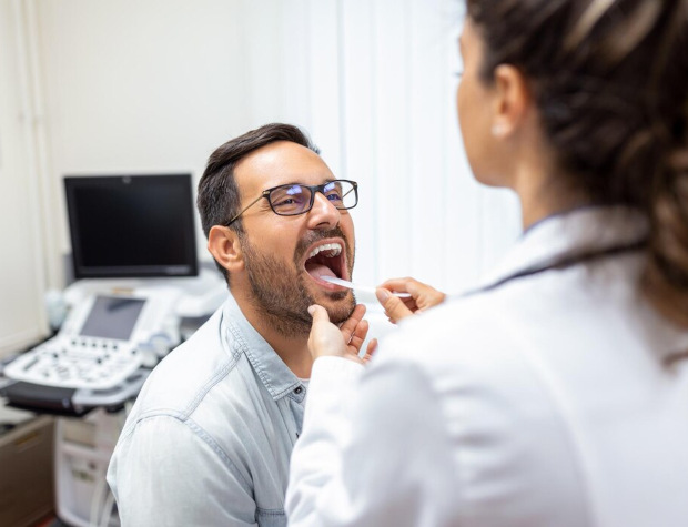
The initial diagnostic stage begins with a consultation with a specialist—a dentist or maxillofacial surgeon. The doctor conducts a questionnaire, identifying the patient's complaints about symptoms and the circumstances of their onset or worsening.
This is followed by a physical examination—a visual assessment of the symmetry of the face and neck, palpation of the salivary glands to determine their size, tenderness, and the presence of masses. An important diagnostic procedure is massage of the gland, assessing the secretion released from its duct.
Instrumental methods play an important role in differential diagnosis, allowing for an objective visual assessment of the glands:
- Ultrasound. Allows for an assessment of the gland's size and structure, and the presence of inflammatory infiltrates, abscesses, stones, or cysts.
- CT or MRI. Prescribed in complex diagnostic cases when it is necessary to clarify the extent of a purulent process, identify small stones, or conduct a differential diagnosis with tumors.
- Sialography is a radiographic contrast study of the ductal system. This method is particularly valuable for diagnosing chronic forms and obstructive processes, as it allows for detailed visualization of duct architecture and the identification of strictures and deformations.
Laboratory methods serve as an important complement, allowing for an overall assessment of the body's condition and identification of the pathogen. A complete blood count reveals nonspecific signs of inflammation: an increased white blood cell count and erythrocyte sedimentation rate, which is especially characteristic of the acute bacterial form. Bacteriological examination of the secretions is performed to accurately identify the pathogenic microflora causing the inflammation and determine its sensitivity to antibiotics, making antibacterial therapy targeted and effective.
Treatment methods
Conservative treatment
Conservative treatments are appropriate for acute uncomplicated and chronic forms of the disease. The doctor's goal is to eliminate the infection, relieve inflammation, reduce swelling, and stimulate the natural flow of saliva.
Drug therapy is selected based on the identified pathogen or the mechanism of pathology development:
- If the inflammation is bacterial in nature, antibiotics are prescribed, the choice of which is based on the results of a bacterial culture.
- If a viral etiology is confirmed, antiviral drugs are used.
- To alleviate the patient's condition, anti-inflammatory and analgesic medications are used to reduce fever, pain, and swelling.
- For chronic processes associated with impaired salivation, salivary stimulants are effective – sialagogues that improve the drainage function of the ducts.
In the early stages, when the inflammation is just beginning to develop and there is no purulent discharge, non-drug treatments can be used. These include:
- A salivary diet, which involves eating foods that stimulate salivation, such as sour fruits, sauerkraut, and fruit drinks.
- Local application of dry heat to improve blood microcirculation.
- Gentle massage of the affected gland along the duct to mechanically remove stagnant secretions.
- Physiotherapy procedures, such as UHF therapy, to dissolve the infiltrate.
- Maintaining an adequate fluid intake to combat dehydration and reduce saliva viscosity.
Please note: Non-drug treatments can only be used after the acute phase has subsided and as prescribed by a doctor.
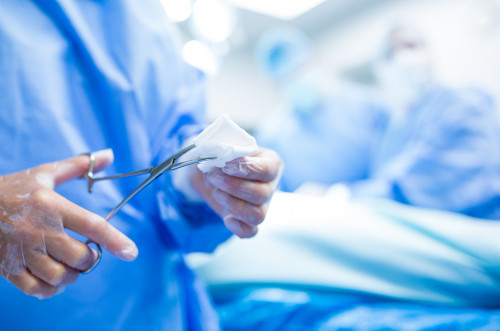
Surgical treatment
Surgical methods are resorted to in cases where conservative approaches are unsuccessful or initially ineffective. Indications for surgery include:
- Formation of a purulent abscess
- Presence of a calculus that does not respond to conservative removal
- Persistent duct narrowing
- Frequent recurrences that significantly reduce quality of life
Modern surgery offers a number of interventions aimed at preserving the organ:
- Incision and drainage of the abscess. This is performed when a purulent cavity forms. The surgeon makes an incision, evacuates the pus, and places a drain to ensure the outflow of the contents.
- Duct bougienage. This procedure is aimed at widening the narrowed excretory duct using special probes.
- Sialendoscopy. This is a minimally invasive endoscopic technique that allows for visual inspection of the ducts from the inside, removal of small stones, or elimination of strictures.
Sialadenectomy, or salivary gland removal, is a last resort used by surgeons in cases of irreversible organ damage, frequent exacerbations, or suspected cancer.
Postoperative patient care
The recovery period after surgery is important for preventing complications and ensuring proper healing. This is facilitated by strict adherence to doctor's recommendations, including:
- Careful care of the postoperative wound with regular dressing changes and antiseptic treatment
- A course of antibiotic therapy to prevent infectious complications
- Following a special diet, which is necessary to protect the surgical site from mechanical and thermal stress
- Regular follow-up examinations by the attending physician to assess healing progress and early detection of possible adverse effects
Recovery times vary. After minimally invasive procedures (abscess incision, sialendoscopy), primary healing takes 7-14 days. Full functional recovery may take several weeks, especially if physical therapy is required. After a sialoadenectomy, the initial postoperative period lasts approximately two weeks, and full adaptation of the body, including compensation of salivary secretion by the remaining glands, can take up to several months.
General information
Possible complications and prognosis for recovery
Delayed or inadequate treatment creates the preconditions for the development of a number of serious complications. The most common consequence is the formation of an abscess – a limited purulent cavity within the glandular tissue, accompanied by increased pain, severe swelling, and requiring mandatory surgical intervention. A more dangerous condition is phlegmon – a diffuse purulent inflammation that has no specific boundaries and can spread to surrounding tissues, posing a threat to life.
Prolonged or recurrent inflammation leads to irreversible structural changes. The ducts narrow, and the functional tissue of the gland is gradually replaced by scar tissue. This causes the disease to become chronic with constant exacerbations. In rare cases, after spontaneous rupture of the abscess, a salivary fistula forms – a pathological channel through which saliva or secretions are released onto the skin.
The prognosis for patients directly depends on the timing of seeking medical attention. With acute sialadenitis diagnosed early and comprehensive conservative therapy, the prognosis is favorable. After a course of therapy, complete recovery and restoration of function occurs.
However, with the development of purulent complications or the chronic course of the disease, the prognosis becomes relatively favorable. In these cases, the goal of treatment is to achieve stable remission and prevent relapses, which requires patient discipline and constant monitoring by a specialist.

Our doctors


This award is given to clinics with the highest ratings according to user ratings, a large number of requests from this site, and in the absence of critical violations.

This award is given to clinics with the highest ratings according to user ratings. It means that the place is known, loved, and definitely worth visiting.

The ProDoctors portal collected 500 thousand reviews, compiled a rating of doctors based on them and awarded the best. We are proud that our doctors are among those awarded.
Make an appointment at a convenient time on the nearest date
Price
Other services

Appointment to the doctor
Reviews


Our clinics

Application “Personal Account K+31”


Basic information about the disease
The salivary glands are an important part of the digestive system, responsible for the production of saliva. The body contains three paired large glandular organs—the parotid, submandibular, and sublingual—as well as hundreds of smaller glands located in the oral mucosa. Malfunction of these structures causes sialoadenitis.
The term "sialoadenitis" refers to a whole group of inflammatory pathologies united by damage to glandular tissue. The inflammatory process leads to swelling, pain, and a significant decrease or cessation of salivary secretion. The large glands, especially the parotid (in this case, the disease is called mumps) and submandibular (submandibulitis), are most vulnerable. This selectivity is due to the anatomical features of their ducts, which are potential gateways for infection.
The physiological role of the salivary glands extends beyond moistening the food bolus. The secretion they produce contains enzymes that initiate the breakdown of carbohydrates in the oral cavity and also has a bactericidal effect. Therefore, inflammation of the salivary gland and the resulting decrease in saliva production negatively impacts the entire digestive process, local immunity, and dental health. A vicious cycle is created: a lack of saliva promotes infection, which further inhibits gland function.