Thoracoscopic surgery

Today, the benefits of minimally invasive surgery are well known and are not in doubt by either doctors or patients. For surgical treatment of various diseases of the abdominal organs , laparoscopic access is currently widely used. Many of laparoscopic surgeries such as cholecystectomy, prostatectomy, hemicolectomy in clinics in the USA, Europe and Asia have become the “gold standard”. The introduction of endoscopic technologies into thoracic surgery was much slower, and for a long time it was limited to performing small diagnostic operations (biopsies, drainage of the pleural cavity, etc.). This is due to the peculiarities of the anatomy of the chest wall (rigid costal framework, limits the degree of freedom of manipulation with instruments), the complex anatomy of the lungs and mediastinum (the presence of large vessels, heart contractions, etc.), as well as the absence of one operation, such as laparoscopic cholecystectomy, which allowed work out endoscopic techniques in detail.

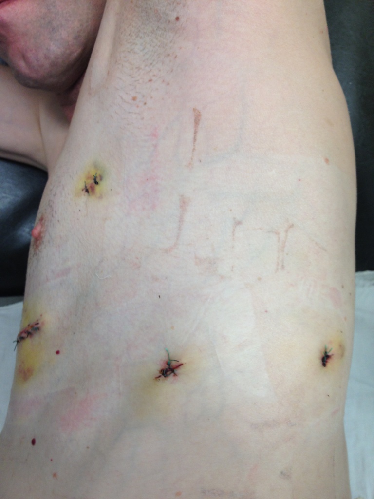
Operations on the lungs and organs of the mediastinum are complex and require a high concentration of attention from the surgeon , detailed knowledge of the anatomy and the exact consistent execution of all stages of the operation. The accumulation of extensive experience in “open” thoracic surgery, the emergence of new instruments and devices for tissue separation has allowed to expand indications for thoracoscopic operations. And today, in the early stages of peripheral lung cancer (a tumor no larger than 5 cm in diameter, without metastases in the lymph nodes), radical thoracoscopic lobectomy (removal of the lobe of the lung) has become the “gold standard”. The same can be said about thoracoscopic thimectomy, which is performed for diseases of the thymus gland through punctures 5-10 mm in size. It should be noted that during surgery for a malignant tumor, the volume of tissue removed is fully consistent with that with “open” access. Due to the increase and high definition of the image, in some cases, it is possible to perform a more thorough audit of the operation area and remove the affected tissue.

The thoracoscopic technique has even greater advantages compared to laparoscopy , since it allows you to avoid blood loss and injury to the intercostal nerves, significantly reduce the severity of postoperative pain, and, in some cases, completely avoid it. The use of ultra-thin instruments of 3 and 5 mm and the finest flexible optics can significantly reduce the patient’s stay in the clinic (up to 2-4 days, compared with 10-12 days after “open” operations), achieve a good cosmetic effect (3 incisions of 5 mm each) and one 4 cm, to remove the tumor or part of the lung) and provide early medical and social rehabilitation.

In the Department of Surgery Clinic + 31 perform thoracoscopic operations for the following reasons:

  • Focal lung lesions, pleura
  • Mediastinal Tumors
  • Peripheral lung cancer
  • Metastatic lung lesion (single metastases)
  • Treatment of spontaneous pneumothorax
  • Thymus disease

Benefits of Thoracoscopic Access

  • Minimum invasiveness: during the operation, retractors that traumatize the intercostal nerves are not used, tools with a diameter of 3, 5 and 12 mm are used. A maximum incision of up to 4 cm is performed at the end of the operation to remove a tumor, cyst, or part of the lung. To visualize the surgical field, special thoracoscopes with a flexible end are used, which allows not changing the position of the camera during the operation. Changes in the operating field occur by controlling the camera using special buttons. Soft and anatomical (corresponding to the shape of the intercostal space) trocars are used.
  • Minimum invasiveness: during thoracoscopic surgery, there is no intersection of a large array of muscles of the lateral surface of the chest, which ensures high quality of life and the absence of deformation of sutured soft tissues. Port installation locations are sutured with self-absorbable threads.
  • Minimal bleeding: accurate allocation of all anatomical structures, the use of modern stapling staplers and hemostasis systems can significantly reduce intraoperative blood loss to 50-100ml.
  • Cosmetic effect: the field of operation remains thin scars, which become almost invisible within 6-8 months. The largest 4cm incision is located in women under the mammary gland.
  • Short rehabilitation and recovery in the postoperative period compared with abdominal surgery.

In Clinic + 31, the majority of diagnostic and therapeutic procedures on the lungs and organs of the mediastinum are performed by thoracoscopic access (in the absence of contraindications). Used equipment from leading manufacturers. Clinic specialists underwent training and internship in thoracoscopic surgery in leading clinics in France and Belgium. The leading specialist in this field is Pavel Kononets (candidate of medical sciences, oncologist surgeon, thoracic surgeon).

Service record


Specialists

All specialists
Udin
Oleg Ivanovich

Deputy chief physician for surgery, surgeon

PhD

Korolev
Sergei Vladimirovich

Deputy chief physician for medical affairs, surgeon, oncologist

PhD

Shapovalyants
Sergei Georgievich

Chief Consultant in Surgery, Surgeon

Tsvetkov
Vitaly Olegovich

Surgeon

Doctor of Sciences, PhD

Malygin
Sergey Evgenyevich

Oncologist-mammologist, surgeon

Kovylov
Aleksey Olegovich

Specialist in wounds and wound infections, diabetic foot doctor

Kim
Ilya Viktorovich

Surgeon

PhD, research fellow

Kovalenko
Yuri Alekseevich

Surgeon

Doctor of Sciences, PhD

Ivanchik
Inga Yakovlevna

Surgeon, phlebologist

PhD

Shpilevoy
Nikolay Yurievich

Cardiovascular surgeon, surgeon, ultrasound specialist

PhD

Natalinov
Ruslan Viktorovich

Surgeon, ultrasound diagnostics doctor