Diagnosis and treatment of hernia of the white line of the abdomen
Hernia of the white line of the abdomen is a disease caused by a defect in the abdominal wall with the subsequent formation of a hernial sac and its protrusion into the resulting gap between the muscles in the abdomen.
To prevent complications, the pathological condition requires immediate treatment. Doctors of the surgery department of the K+31 clinic (Moscow) perform hernioplasty operations using modern minimally invasive technologies.

specialists

equipment

treatment


Causes of occurrence
Stretching of the white line of the abdomen can occur due to congenital dysplasia of connective tissue or as a result of diseases in which metabolism is disrupted (diabetes, autoimmune pathologies).
With a weakened white line, a hernia can form due to increased pressure inside the abdominal cavity.
Provoking factors include:
- Increased loads associated with lifting weights (heavy physical labor, sports)
- Pregnancy
- "Apple" obesity (excessive accumulation of fat in the upper body) with insufficient physical activity
- Prolonged cough in chronic lung diseases
- Accumulation of free fluid in the peritoneum
- Past injuries or operations in the center of the abdomen
- Frequent constipation, which requires constant abdominal strain
Pathology of the white line of the abdomen occurs after 40 years, more often in women. In children, it can occur with prolonged and heart-rending crying.
Symptoms
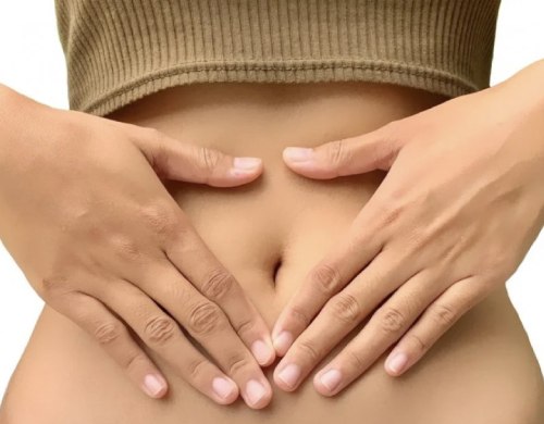
At the early stage of the disease, the patient does not experience pain. A small bulge may appear on the abdomen, in its central part. As the hernial sac comes out, the bulge takes on a round or oval shape, and its diameter can reach up to 12 cm.
The main symptoms of the pathology:
- The bulge occurs when coughing or physical exertion, causing discomfort or pain, in the lying position the bulge decreases or disappears completely
- At moments of short-term strangulation, pain and nausea (with vomiting) occur
At the early stage, the patient can independently reduce the hernia.
In advanced cases, the strangulation of the internal organs that have come out in the hernial orifice is manifested by sharp, burning pain, increasing nausea, constipation, and the inability to independently reposition the formation. Against the background of symptoms of intoxication (cold sweat, fever, dizziness), a feeling of a "bloated abdomen" appears, but gases do not pass. Traces of blood in the feces are possible during defecation.
Important! If signs of strangulation of the hernia appear, you must quickly consult a doctor. To exclude irreversible consequences, an emergency operation is performed.
Classification and stages of hernia development
The pathology is classified by:
- The cause of occurrence - congenital and acquired
- The nature of the course - reducible and irreducible
- The place of occurrence - periumbilical, supraumbilical and subumbilical
- The number of defects - single and multiple
The hernia goes through three stages in the process of its formation:
- A preperitoneal lipoma appears against the background of relaxation and "divergence" of the muscular-aponeurotic layer of the peritoneum, forming a hernial orifice. There are no external manifestations, but palpation may cause pain
- A hernial sac is formed from fatty tissue and peritoneal tissue. When straining, a protrusion on the abdomen periodically forms, which disappears on its own during rest
- Parts of the internal organs (usually the intestines, omentum or stomach walls, and in pregnant women in the last trimester, the uterus) come out into the hernial sac. The volume of the protrusion increases, pain becomes more frequent. At this stage, compression of the outgoing organs in the hernial orifice is possible
Surgical intervention to remove a hernia of the white line of the abdomen is necessary at stages 2 and 3 of the pathology.
Pathogenesis of hernia of the white line of the abdomen
The main factors in the development of hernia include a violation of the structure of the connective tissue, when an imbalance occurs between the amount of mature and immature collagen fibers. Dysplasia develops if there is much more immature collagen (type 3) in the connective tissue than mature (type 1). In this case, a thinner and weaker layer of the muscular-aponeurotic system is formed in the abdominal wall.
A weakened white line of the abdomen does not provide the necessary support to the internal organs between the rectus abdominis muscles, and with an increase in intra-abdominal pressure, ruptures are formed.
In children, the muscular-aponeurotic layer is weaker than in adults. Therefore, against the background of severe chronic diseases with prolonged cough or constipation, the white line stretches faster, forming a hernia.
In adults, the maturation of collagen fibers can be hindered by an excess layer of fatty tissue on the abdomen and weakness of the abdominal muscles. This can also be affected by pregnancy (multiple or frequently repeated). Any increased load immediately affects the "weak spots" and a hernia comes out through the stretched wall.
Diagnostics of a hernia of the white line of the abdomen
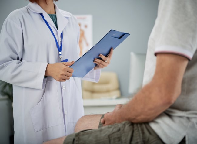
During the initial consultation, the specialist examines and palpates the area of the protrusion, taps it. The patient needs to strain so that it is possible to assess the degree of protrusion of the hernial sac and the possibility of its reduction.
For differential diagnostics, the following is performed:
- Ultrasound of the hernia and its contents (if large)
- Ultrasound of the abdominal cavity
- X-ray
- Irrigoscopy
- Herniography with contrast
MRI is prescribed in complicated cases (with recurrent hernia, obesity or severe pain in the abdominal wall without visible detection of the hernia), when it is necessary to assess the condition of the surrounding tissues. These diagnostic methods allow you to confirm the diagnosis and differentiate the pathology from others with similar symptoms.
Treatment of hernia of the white line of the abdomen
Children under 5 years of age are prescribed conservative therapy and monitoring of the hernia condition. Most often, wearing a bandage is recommended and physiotherapy is prescribed.
For adults, the main method of treatment is surgery to remove a hernia of the white line of the abdomen. The main goal of surgical intervention is to push the internal organs back into the abdominal cavity and remove the hernial sac.
Treatment methods
There are two main ways to eliminate the abdominal wall defect:
- Hernioplasty with tension (fascial-aponeurotic plastic surgery using muscle tissue). The hernial orifice is eliminated by tightening the surrounding muscle tissue and applying sutures. During the operation, the doctor makes a vertical incision and tensions the patient's own tissues. Hernioplasty involves long-term rehabilitation with the exclusion of any loads. It is prescribed for small hernias at an early stage in the absence of complications
- Hernioplasty without tension (artificial materials are used). To eliminate the abdominal wall defect, a mesh or endoprosthesis is applied. The implant is applied to the area of the hernial orifice according to the "flap" principle and it holds the surrounding tissues. After the operation, the mesh material or endoprosthesis is overgrown with muscle fibers, creating a strong frame. Hernioplasty reduces the likelihood of relapse to a minimum. Used for large diameter hernias
The operation is performed using an open or laparoscopic method. Cavity intervention is recommended for frequent relapses in case of complications.
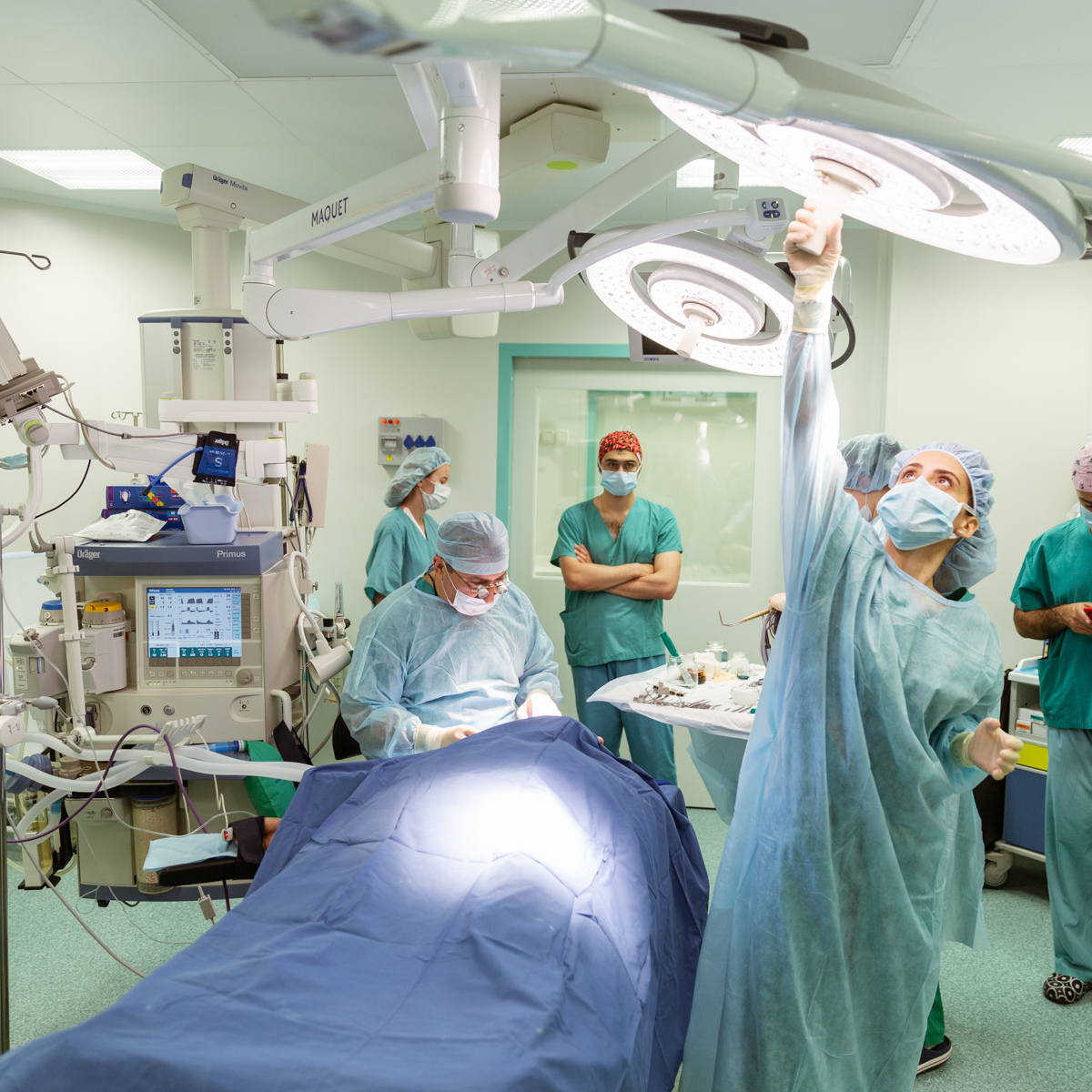
Surgical treatment
It can be performed on a planned or emergency basis. In the first case, the doctor prescribes the operation for a certain time in the hospital and selects one of the types of hernioplasty based on the condition of the surrounding abdominal tissues.
Emergency hospitalization is necessary if there are complications. In this case, preparation may take 1 day, and the surgical intervention is performed in an open manner.
Algorithm for performing hernioplasty:
- Administration of anesthesia. Treatment of the surgical area with an antiseptic
- Providing access to the hernia through an external tissue incision with a scalpel or a laparoscope
- Releasing the internal organs from the hernial sac and reducing them to an anatomical position
- Excision and removal of the hernial sac
- Elimination of the abdominal wall defect using a tension or non-tension method
- Application of external sutures
- Disinfection of the wound and fixation of a sterile dressing
In case of strangulation of the hernia, the doctor removes the hernial sac along with its contents, excises the damaged tissue and restores the integrity of the internal organs. In the presence of peritonitis, the abdominal cavity is washed and the hernial orifice is then sutured.
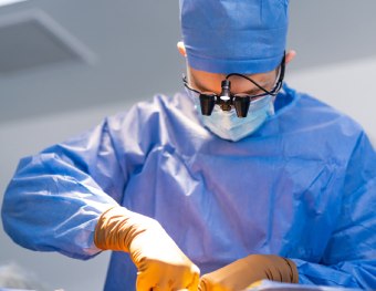
Endovideosurgical methods of hernioplasty
Modern low-traumatic methods of surgical intervention include laparoscopic hernioplasty. During the operation, the doctor does not make a cavity incision. Endoscopic equipment with built-in optics, a camera, lighting and microsurgical instruments are installed into the peritoneal cavity through small punctures.
Using the endovideosurgical method, the surgeon performs the operation, suturing the patient's own tissues or installing a mesh endoprosthesis.
Advantages of the method:
- No large skin incisions, small (5 mm) scars remain after the operation at the puncture sites of the abdominal cavity
- A more detailed overview of the tissue condition through a camera that displays an image of the internal area of the operation magnified tens of times on an external screen
- Fast rehabilitation
- Minimal risk of relapse
However, laparoscopic surgeries also have contraindications. They are not performed in the event of complications, tissue necrosis and peritonitis, requiring emergency intervention. The method is also not suitable for repeated surgery, in case of relapse after a previous intervention in the same place.
General information
Prognosis and prevention
If you see a doctor in a timely manner and the hernia is eliminated, the prognosis for recovery is favorable. The likelihood of relapse is associated with the condition of the tissues, the method of surgery and the quality of rehabilitation. With laparoscopic hernioplasty, relapse is up to 5%. In the case of abdominal operations, especially when using the tension method, relapse can occur in 5-15% of cases.
To avoid hernia or recurrence in the postoperative period, it is necessary to adhere to the following recommendations:
- Eliminate the cause of the pathology - cure chronic diseases of the gastrointestinal tract and lungs
- Avoid coughing, sneezing and lifting heavy objects for 3 months after surgery
- To prevent constipation, follow a high-fiber diet
- Be physically active, but without lifting heavy objects or straining the abdominal muscles
- Monitor your body weight
- Wear a bandage for no more than 1 month, since prolonged immobilization weakens the muscles even more
To avoid hernia formation, you should train your abdominal muscles. In order to prevent complications, you should not neglect a hernia until it spontaneously infringes. You should see a doctor for examination and treatment as soon as the pathology is detected.


FAQ
How long does it take to recover from a hernia of the white line of the abdomen?
On average, rehabilitation after surgery is 1-2 months. During this time, the wound heals, the functions of the internal organs are restored. The first 2-3 weeks, it is necessary to monitor the condition of the sutures, exclude constipation and cough. For 3 months after the operation, you cannot lift anything heavy (more than 5 kg).
What kind of anesthesia is used?
General anesthesia is used for hernia removal.
How often does a relapse of a hernia of the white line of the abdomen occur?
If the doctor's recommendations are followed during the rehabilitation period after surgery, a relapse is possible in 5% of cases. The hernia may reappear with early physical activity or with severe pathologies of the connective tissue.
Sources and literature
- Grigoryev S.G., Bratiychuk A.N., Krivoshekov E.P., Grigorieva T.S. - Hernioplasty Technologies in Same-Day Surgery, 2005
- Zhebrovsky V.V., Mohammed Tom Elbashir. Surgery of Abdominal Hernias and Eventrations. Simferopol, 2012
- Gorbunov N.S., Samotesov P.A., Petruschko S.I. "Structure of the White Abdomen Linea." Siberian Medical Review, Vol. 69, No. 3, 2021
Our doctors


This award is given to clinics with the highest ratings according to user ratings, a large number of requests from this site, and in the absence of critical violations.

This award is given to clinics with the highest ratings according to user ratings. It means that the place is known, loved, and definitely worth visiting.

The ProDoctors portal collected 500 thousand reviews, compiled a rating of doctors based on them and awarded the best. We are proud that our doctors are among those awarded.
Make an appointment at a convenient time on the nearest date
Price
Other services

Appointment to the doctor
Reviews


Our clinics

Application “Personal Account K+31”


General information
The white line of the abdomen is a plexus of tendons and fascia connected to the muscular aponeurotic layer and located on the sides of the rectus abdominis muscles. It looks like a vertical stripe running from the sternum to the pubis through the navel. It got its name because of its characteristic white color.
This place in the abdominal wall is relatively weak. Under the influence of various internal and external factors, it can "diverge". Part of the internal organs (usually intestinal segments, adipose tissue, omentum) penetrates into the resulting gap, thus forming a hernial sac. Most often, a hernia forms in the upper abdomen, less often near the umbilical ring or below it.
Compared to umbilical and inguinal, this pathology is much less common (2-4% of cases). The danger is the strangulation of the hernial sac with subsequent tissue necrosis, peritonitis and sepsis. The pathology is treated only by surgery.