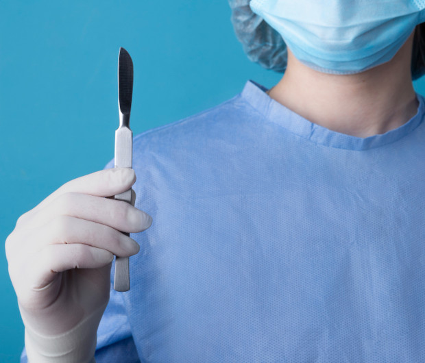
Inguinal Hernia Surgery
An inguinal hernia is a condition in which abdominal organs protrude beyond the hernia. This manifests as a characteristic bulge in the groin. It is often associated with discomfort and pain of varying intensity. A radical treatment method that can correct the defect and prevent the risk of serious complications such as strangulation is surgical intervention – hernioplasty.
Modern medicine offers effective and minimally invasive techniques that ensure reliable closure of the hernial orifice and rapid recovery. At the K+31 Clinic in Moscow, hernia surgeries are performed using advanced techniques, achieving excellent results.

specialists

equipment

treatment


Causes and symptoms
The development of a hernia is the result of a combination of two factors: a weak spot in the musculoaponeurotic framework and a regular or sudden increase in intra-abdominal pressure. The clinical picture can range from virtually asymptomatic to severe pain and the appearance of a clearly visible bulge, requiring immediate medical attention.
Causes of the development of pathology in men
Men experience this condition significantly more often than women, which is directly related to their anatomical features. The inguinal canal in men is wider, shorter, and less supported by muscle and tendon fibers. Furthermore, it serves as a natural passage for the spermatic cord, which poses an additional risk.
The main causes and factors that contribute to hernia formation include:
- Connective tissue weakness. This is a congenital or acquired condition in which the muscle fibers that make up the abdominal wall are not strong enough. This condition, known as connective tissue dysplasia, often leads to hernia development even without significant physical activity.
- Age-related changes. Over time, muscles and tendons naturally weaken and thin, reducing their elasticity. Therefore, inguinal hernias are often diagnosed in middle-aged and older men.
- Heavy workload. Regular heavy lifting, strength training, or work involving physical labor create constant increased pressure on the anterior abdominal wall, which gradually pushes organs through weak spots.
- Chronic cough. A persistent, severe cough, which accompanies conditions such as bronchitis, COPD, or asthma, greatly increases intra-abdominal pressure.
- Gastrointestinal disorders. Chronic constipation requires regular straining during bowel movements, which also contributes to the formation of a hernia.
- Overweight. Obesity places constant additional strain on the abdominal muscles, simultaneously contributing to the weakening of the muscular corset.
Clinical manifestations
Symptoms depend on the stage of development and size of the hernia. Typically, they include:
- Visible bulge. The most characteristic sign is the appearance of a soft, elastic swelling. In the early stages, it may be barely noticeable and periodically disappear, returning to the abdominal cavity on its own.
- Discomfort and nagging pain. A feeling of heaviness, pressure, or aching in the lower abdomen and groin are common symptoms of a hernia. The discomfort usually intensifies towards the end of the day, after physical activity, and subsides with rest. Many report a sensation of a foreign body in the groin.
- Symptoms worsen with exertion. A characteristic feature of the pathology is an increase in the protrusion and increased pain with any activity that increases intra-abdominal pressure: coughing, sneezing, straining, or lifting.
Particular attention should be paid to symptoms of strangulation—a life-threatening complication in which organs are compressed within the hernial orifice, disrupting their blood supply. This condition requires emergency surgical intervention.
Warning signs:
- Sharp, sudden pain in the hernia area that does not subside with rest.
- Inability to reduce a hernia that was previously easily reducible.
- Redness of the skin over the hernia sac, localized fever.
- Nausea, vomiting, lack of bowel movements, and lack of gas.
If any of these symptoms occur, seek emergency medical attention immediately.
Excision surgery: indications and contraindications

The decision to perform surgical intervention is based on a careful assessment of the benefit-risk ratio for each individual patient. In modern surgery, surgery is the only radical treatment method that can correct the defect and prevent dangerous complications.
Elective surgical treatment is indicated for all patients with a confirmed diagnosis, regardless of the size of the hernia. Even small and asymptomatic hernias are prone to progression, and their planned removal is technically simpler and safer for the patient than emergency surgery in case of complications.
Surgical intervention is strongly recommended as soon as possible in the following cases:
- Risk of strangulation. This can occur suddenly and lead to necrosis of the strangulated organ, peritonitis, and acute intestinal obstruction, which poses a direct threat to life.
- Pain and discomfort. Constant pain, heaviness, pressure, or discomfort in the groin that limits physical activity, disrupts daily life, and reduces quality of life.
- Increasing hernia size. Rapid growth of the hernia or its large size not only creates a significant cosmetic defect but also increases the risk of complications and makes future surgery more technically challenging.
- Irreducibility. If the hernial contents no longer return freely to the abdominal cavity while lying down, this indicates the formation of adhesions, a harbinger of possible strangulation.
Like any surgical procedure, inguinal hernia surgery has its contraindications.
Absolute:
- Acute infectious and inflammatory diseases – intervention is postponed until complete recovery;
- Decompensated diseases of vital organs – severe cardiac, renal, or hepatic failure;
- Recent myocardial infarction or stroke;
- Severe bleeding disorders;
- Terminal stages of oncological diseases.
Relative:
- Compensated chronic diseases – coronary heart disease, hypertension, diabetes mellitus;
- Mild forms of coagulopathies amenable to drug correction;
- Benign tumors that do not pose an immediate risk to life;
- High obesity – however, the patient may Preliminary weight loss may be recommended.
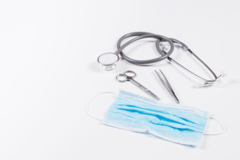
Diagnostics
An accurate diagnosis is a crucial step in determining further treatment. Although the diagnosis often seems obvious, a comprehensive examination is necessary to determine the type and size of the hernia, the condition of the surrounding tissues, and to differentiate it from other groin conditions, such as lymphadenitis, varicocele, and hydrocele. This allows for the selection of the most effective and safe surgical procedure.
Physical examination if pathology is suspected
The first stage of diagnosis is a consultation with a surgeon and a physical examination. The examination begins with a visual assessment, both standing and lying down, which allows for the detection of even minor asymmetries and early detection of a protrusion.
The doctor palpates the inguinal canal using a cough test. To do this, they gently insert a fingertip into the external inguinal ring and ask the patient to strain or cough. When intra-abdominal pressure increases, the surgeon feels a characteristic push with their finger.
Next, the hernia's reducibility is assessed. In the supine position, the doctor can gently push the contents of the hernial sac back into place. This is also informative: easy reducibility is typical of uncomplicated hernias, while failure to do so is a worrying sign and may indicate the presence of adhesions or strangulation.
Advanced instrumental diagnostics
To confirm the diagnosis, clarify the details of the hernia's anatomy, and rule out other pathologies, instrumental methods are used.
- Ultrasound examination of the scrotum and inguinal canals. This is a non-invasive, safe, and highly informative method. With its help, the surgeon visualizes the hernial sac itself and its contents (omentum, intestinal loop), accurately determines the size of the hernial orifice, assesses the condition of the muscles and aponeuroses, and differentiates an inguinal hernia from other formations, such as a cyst or tumor. The examination is performed in real time, including the use of a straining test.
- MRI or CT are typically used in complex diagnostic cases. These methods may be recommended for relapses to assess postoperative anatomy and identify the causes of recurrence, if rare and complex forms of pathology are suspected, such as interstitial hernias, in the presence of large inguinal-scrotal hernias, and also if the clinical picture is unclear and it is necessary to exclude other diseases of the pelvic organs and abdominal cavity.
Preparation for Surgery
Thorough preparation for hernioplasty is a mandatory step that directly impacts the success of the procedure and minimizes the risk of intra- and postoperative complications. A well-organized preoperative process allows for a comprehensive assessment of the patient's health, identification and consideration of possible deviations, and psychological and physical preparation for the upcoming procedure.
Basic Preparation Requirements
The preparatory stage begins with a comprehensive laboratory and instrumental examination, the purpose of which is to assess the body's readiness for anesthesia and surgery. It includes:
- Standard preoperative testing package
- ECG
- Consultations with a general practitioner, anesthesiologist, and, if necessary, specialists such as a gastroenterologist or cardiologist.
- Correction of chronic diseases – achieving a stable condition in pathologies such as arterial hypertension, diabetes, and others.
- Treatment of infection foci – it is necessary to treat caries, chronic tonsillitis, or other sources of inflammation in advance, as they can cause postoperative complications.
Preparation Immediately Before Surgery
Shortly before hernioplasty, the patient must follow a number of important rules:
- Diet. 2-3 days before surgery, it is recommended to avoid foods that cause flatulence and increased gas production. These include legumes, brown bread, cabbage, and carbonated drinks. A light dinner is allowed the night before.
- Fasting break. 8-12 hours before surgery, you must completely abstain from all food intake, and 2-3 hours before surgery, you must abstain from drinking water. This strict rule is aimed at preventing aspiration – the aspiration of stomach contents into the respiratory tract during anesthesia due to vomiting.
- Hygiene procedures. A shower is recommended on the morning of surgery. The surgical site is shaved directly in the clinic by medical staff using sterile instruments. Shaving the day before is not recommended due to the high risk of microtrauma and skin irritation, which can become a gateway for infection.
- Cleansing enema. May be recommended for patients prone to constipation or when planning certain types of anesthesia to empty the bowels and prevent postoperative bowel obstruction. Not required for everyone.
- Jewelry removal. Before being transferred to the operating room, all jewelry, as well as removable dentures and contact lenses, must be removed. This is due to safety requirements.
Surgical Methods
Several hernia repair techniques are currently practiced, divided into two broad groups: open and laparoscopic. The choice of technique depends on the size and type of hernia, the patient's age, and the presence of comorbidities. The main goal of any intervention is not only to eliminate the protrusion but also to strengthen the weakened wall to prevent recurrence.
Laparoscopy
Laparoscopic hernioplasty is a minimally invasive procedure performed through several small incisions in the anterior abdominal wall. Carbon dioxide is pumped into the abdominal cavity to create a working space, after which instruments and a laparoscope (a video camera with a light source) are inserted through the incisions. The surgeon performs the procedure using the camera image displayed on a monitor as a guide.
There are two main techniques for performing laparoscopic surgery:
- Transabdominal preperitoneal hernioplasty (TAPP). The surgeon operates in the abdominal cavity, making an incision into the peritoneum over the hernia defect. After the mesh implant is placed and secured in the preperitoneal space, the peritoneum is sutured, isolating the mesh from the internal organs.
Total extraperitoneal hernioplasty (TEP). The hernia is approached without entering the abdominal cavity. The surgeon creates a space between the peritoneum and abdominal muscles, where the mesh is placed. This laparoscopic technique is considered less invasive than TAPP.
The laparoscopic technique has several advantages. The main ones are:
- Minimal trauma – the absence of a large incision minimizes tissue damage.
- Less pain compared to open surgeries.
- Cosmetic effect – barely noticeable scars remain after healing.
- Reduced recovery period – quick return to active life and work.
Tension and tension-free hernioplasty.
These are open surgical methods that access the inguinal canal through a 4-6 cm skin incision in the groin area.
Tension hernioplasty is considered an outdated method. The essence of the technique is to eliminate the hernial defect by tightening and suturing the patient's own tissues at the hernial orifice without the use of implants. This creates significant tissue tension, which is the main drawback of this method. High tension leads to severe pain, prolonged rehabilitation, and, most importantly, a high recurrence rate (up to 10-15%), as the tissues often cannot withstand the strain.
Tension-free hernioplasty is the modern standard for open surgery. With this technique, the hernial orifice is closed not by contracting the patient's own tissues, but by implanting a special mesh prosthesis made of synthetic material. The mesh is placed in the defect area and reliably strengthens the posterior wall of the inguinal canal. Over time, it grows into the patient's own tissues, forming a strong anatomical barrier.
Advantages of the tension-free technique:
- Absence of tissue tension, which significantly reduces the intensity of postoperative pain.
- Extremely low recurrence rate (less than 1-2%).
- Shorter and easier recovery period compared to tension-based techniques.
- The surgery can be performed under local anesthesia in patients at high risk for general anesthesia.
Methods of performing surgical intervention
Several hernia repair techniques are currently practiced, divided into two broad groups: open and laparoscopic. The choice of technique depends on the size and type of hernia, the patient's age, and the presence of comorbidities. The primary goal of any procedure is not only to eliminate the hernia but also to strengthen the weakened wall to prevent recurrence.
Laparoscopy
Laparoscopic hernioplasty is a minimally invasive procedure performed through several small incisions in the anterior abdominal wall. Carbon dioxide is injected into the abdominal cavity to create a working space, after which instruments and a laparoscope (a video camera with a light source) are inserted through the incisions. The surgeon performs the procedure using the camera image displayed on a monitor as a guide.
There are two main techniques for performing laparoscopic surgery:
- Transabdominal preperitoneal hernioplasty (TAPP). The surgeon operates in the abdominal cavity, incising the peritoneum over the hernial defect. After inserting and securing the mesh implant in the preperitoneal space, the peritoneum is sutured, isolating the mesh from the internal organs.
- Total extraperitoneal hernioplasty (TEP). The hernia is accessed without entering the abdominal cavity. The surgeon creates a space between the peritoneum and abdominal muscles, where the mesh is placed. This laparoscopic method is considered less invasive than TAPP.
The laparoscopic method has several advantages. The main ones are:
- Minimal invasiveness – the absence of a large incision minimizes tissue damage.
- Less painful than open surgery.
- Cosmetic effect – barely noticeable scars remain after healing.
- Shorter recovery period – quick return to active life and work.
Tension and non-tension hernioplasty
These are open surgical methods that access the inguinal canal through a 4-6 cm skin incision in the groin area.
Tension-assisted hernioplasty is considered an outdated method. The technique involves eliminating the hernia defect by contracting and suturing the patient's own tissues at the hernial orifice without the use of implants. This creates significant tissue tension, which is the main drawback of this method. High tension leads to severe pain, prolonged rehabilitation, and, most importantly, a high recurrence rate (up to 10-15%), as the tissues often cannot withstand the strain.
Tension-free hernioplasty is the modern standard for open surgery. With this technique, the hernial orifice is closed not by contracting the patient's own tissues, but by implanting a special mesh prosthesis made of synthetic material. The mesh is placed in the defect area and reliably strengthens the posterior wall of the inguinal canal. Over time, it grows with the patient's own tissue, forming a strong anatomical barrier.
Advantages of the tension-free technique:
- Absence of tissue tension, which significantly reduces the intensity of postoperative pain.
- Extremely low recurrence rate (less than 1-2%).
- Shorter and easier recovery period compared to tension techniques.
- The surgery can be performed under local anesthesia in patients at high risk for general anesthesia.
How does the operation proceed?
The surgical treatment of an inguinal hernia is always strictly organized and follows a pre-planned procedure. It involves a team of specialists: a surgeon, an anesthesiologist, and scrub nurses.
-
Hospitalization and preoperative preparation. The patient is admitted to the medical center on the day of surgery or the day before. In the preoperative room, they change into special clothing. Medical staff monitor blood pressure and temperature. A peripheral venous catheter is inserted for infusions and medication administration.
-
Anesthesia Selection and Administration. The type of anesthesia is selected individually during a preliminary consultation with an anesthesiologist and depends on the surgical method, the patient's health, and their preferences. Local, spinal, and general endotracheal anesthesia are available.
-
The surgical procedure itself. Depending on the chosen technique, the surgeon makes an incision or punctures and excises the hernial sac, then sutures the tissue. Open tension-free hernioplasty typically takes 30 to 60 minutes. Laparoscopic surgery, being more technically complex, can last 45 to 90 minutes.
-
Recovery from anesthesia and transfer to the ward. After the surgery, the anesthesia wears off, and the patient gradually awakens. Doctors monitor the patient's blood pressure, pulse, and oxygen saturation. After the patient's condition stabilizes, they are transferred to the inpatient ward, where they continue their recovery.
General information
Postoperative Period
The surgery itself is only half the battle. Strict adherence to doctor's recommendations during the postoperative period largely determines the speed of recovery and long-term results.
Postoperative Rehabilitation
During the recovery period, special attention is paid to three aspects: returning to physical activity, managing nutrition, and caring for sutures.
One of the main advantages of modern techniques is the ability to move within a few hours after surgery. Getting out of bed and walking around the ward and hallway on the day of surgery or the following morning is necessary to prevent thromboembolic complications and pneumonia, as well as to stimulate bowel function.
Despite early activation, strict restrictions remain. During the first month, lifting weights over 3-5 kg, doing abdominal exercises, engaging in heavy physical labor, and sports are strictly prohibited. After 1-3 months, depending on the surgical method and individual circumstances, you can gradually return to your normal activities after consulting with your surgeon.
The main goal of following a diet is to prevent constipation and excess gas, which increase intra-abdominal pressure and put unnecessary strain on the surgical site. Recommended:
- Drink plenty of water
- Eat fiber-rich foods: boiled cereals, stewed vegetables, fruit purees
- Avoid foods that cause flatulence, as well as spicy, smoked foods, and alcohol.
If the wound is sutured with non-absorbable material, the sutures are removed on the 7-10th day. Until then, keep the postoperative dressing, if present, clean and dry. Allowing the incision to get wet is only permitted with your doctor's permission. Antiseptic treatment of sutures is performed as recommended by the surgeon.


Our advantages and specialists
The K+31 Clinic purposefully creates an environment in which modern medical technologies are combined with an individualized approach to each patient, ensuring a successful treatment outcome.
We offer:
- Expert surgeons. Surgeries are performed by highly qualified hernia surgeons who specialize in hernia treatment and are proficient in all modern techniques.
- Advanced department equipment. We use modern surgical equipment from leading global manufacturers, high-quality mesh implants, and consumables.
- Individualized treatment plan. No two patients are alike, so we don't offer cookie-cutter solutions. The surgical method, type of anesthesia, and postoperative care are chosen strictly on a case-by-case basis.
- High level of service. Treatment takes place in a modern, comfortable, fully equipped hospital. We offer comfortable rooms, organized dietary meals, and 24-hour medical supervision.
- Affordable prices with the highest quality. Our goal is to ensure that the cost of surgery, using high-quality implants and a comfortable hospital stay, remains predictable and reasonable.
By contacting the K+31 clinic, you entrust your health to a team of professionals who utilize all the possibilities of modern medicine to ensure your speedy return to a full, active life.

Our doctors


This award is given to clinics with the highest ratings according to user ratings, a large number of requests from this site, and in the absence of critical violations.

This award is given to clinics with the highest ratings according to user ratings. It means that the place is known, loved, and definitely worth visiting.

The ProDoctors portal collected 500 thousand reviews, compiled a rating of doctors based on them and awarded the best. We are proud that our doctors are among those awarded.
Make an appointment at a convenient time on the nearest date
Price
Other services

Appointment to the doctor
Reviews


Our clinics

Application “Personal Account K+31”


What is an inguinal hernia?
This is a condition in which loops of small intestine or omentum protrude beyond the anatomical boundaries of the abdominal cavity through a weakened or dilated inguinal canal. Essentially, this hernia is a pocket formed by the peritoneum that, under pressure from intra-abdominal forces, penetrates the inguinal canal, creating a visible lump in the groin area.
The inguinal canal is a narrow space, 4-6 cm long, located in the lower part of the abdominal wall. Normally, it serves as a natural tunnel for the passage of the spermatic cord or the round ligament of the uterus. Its walls are formed by muscles and aponeuroses, which normally securely hold the organs in place. However, when these structures weaken, their function is impaired, creating the preconditions for hernia formation.
Inguinal hernias are classified according to two main criteria: anatomical location and origin.
Anatomically, there are two types:
By origin, hernias are divided into: