Ultrasound (ultrasound diagnostics)

Advantages and features of ultrasound
- High information content about the condition of internal organs, tissues, tendons, cartilage, ligaments, blood vessels of the head
- The ability to determine changes in organ structures and find tumors in the early stages of development
- Safety and absence of absolute contraindications
- Ability to assess the condition of the fetus, newborns and infants
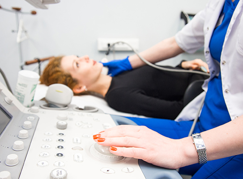
- Non-invasive and painless (the sensor is placed on the surface of the skin above the area under study)
- The ability to assess the dynamics of the development of the disease without harm to the body
- The protocol is done immediately during the procedure
- Availability of modern, professional ultrasound scanners for accurate diagnostics in the medical center
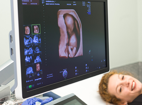
According to the International Society of Ultrasound in Obstetrics and Gynecology (ISUOG), the best ultrasound diagnostic system for performing ultrasound during pregnancy in all trimesters is the General Electric Voluson E10, which our clinic also has.
We offer expectant mothers ultrasound examination of the fetus at various stages of pregnancy. Using our equipment, it is possible to conduct screening to assess intrauterine development.
Doppler ultrasound of the uteroplacental and fetal blood flow has important diagnostic value in the group of pregnant women at high perinatal risk.
Ultrasound is quite informative for multiple pregnancies, various pathologies, and late pregnancy. The research method allows us to identify pathological changes and anomalies in fetal development.
Timely detection of violations makes it possible to prevent the development of severe complications of pregnancy and develop tactics for managing a pregnant woman.
Shear wave liver elastometry is performed on the Philips Affinity 70 expert-class ultrasound machine in the clinic. Diffuse liver disease is one of the biggest problems in the world. The etiology of this type of disease is diverse:
- Viral hepatitis (hepatitis B or C).
- Fatty infiltration of the liver of alcoholic and non-alcoholic origin.
- Autoimmune hepatitis.
- Medication-related complications
- Primary biliary cirrhosis and other less common pathological conditions and diseases.
Our clinic uses the recommendations developed by the Russian Association of Ultrasound Diagnostics in Medicine and the Department of Ultrasound Diagnostics of the University of Pavia (Italy) to assess liver stiffness mainly against the background of viral hepatitis C. Point shear wave elastography is based on measuring the speed of propagation of a shear wave and allows a specialist to obtain a digital value of liver stiffness, which can be correlated with various rating scales, including the METAVIR scale.
Services
Ultrasound of the kidneys and adrenal glands
Ultrasound of the bladder, ureters and urinary tract
Ultrasound of the penis and testicles
Prostate ultrasound (transabdominal and transrectal)
Pediatric ultrasound doctor
Ultrasound of the scrotum
Biopsy under ultrasound guidance Ultrasound gynecological Ultrasound in urologyOur doctors


This award is given to clinics with the highest ratings according to user ratings, a large number of requests from this site, and in the absence of critical violations.

This award is given to clinics with the highest ratings according to user ratings. It means that the place is known, loved, and definitely worth visiting.

The ProDoctors portal collected 500 thousand reviews, compiled a rating of doctors based on them and awarded the best. We are proud that our doctors are among those awarded.
Make an appointment at a convenient time on the nearest date
Price

To get a consultation
Answers to popular questions
What technologies do K+31 doctors use for ultrasound?
Clinic K+31 has stationary and portable ultrasound devices of an expert class from leading manufacturers of ultrasound equipment (Toshiba, General Electric, Philips, Hitachi):
- Wide range of diagnostic capabilities
- Excellent degree of visualization of the structure of organs and tissues
- Doppler research programs
- Panoramic scanning
- Building a volumetric image in real time
The premium ultrasound devices Canon i800, Toshiba Artida, Toshiba Aplio 500 installed in the clinic include innovative ultrasound imaging technologies and have a number of unique capabilities that allow obtaining grey-scale images of unprecedented quality with exceptionally detailed elaboration of tissue and organ structures.
New possibilities for computer processing of ultrasound images in three-dimensional (3D-4D) mode on the latest generation devices allow you to create a spatial picture of the structure of an organ, vessel, or pathological focus.
Using our modern equipment, it is possible to diagnose pathologies of the urinary system and reproductive system.
What organs and systems can be checked by ultrasound in K+31?
K+31 carries out almost all currently existing ultrasound research methods:
- Brain
- Abdominal organs
- Kidneys and adrenal glands
- Liver
- Thyroid
- Mammary glands
- Salivary glands
- Lymph nodes
- Lungs
- Pleural cavities
- Orbits of the eye and sockets
- Pelvic organs in men and women
- Bladder
- Organs of the scrotum
- Prostate gland (prostate)
- Soft tissues and joints
- Abdominal cavity
- Gall bladder
- Retroperitoneal space
- Pancreas
- Spleen
- Lower limbs
- Upper limbs
- Heart (EchoCG)
- Vessels
- Triplex and duplex scanning of great vessels and veins, arteries of internal organs
- Scanning of the brachiocephalic arteries (with color Doppler mapping of blood flow, with rotational tests)
- Doppler echocardiography and stress echocardiography, transesophageal echocardiography
- Transcranial Dopplerography (TCDG)
- Ultrasound diagnostics during pregnancy in all trimesters
- Transvaginal examination in the first trimester of the uterus, cervix
- Dopplerography of uteroplacental and fetal blood flow
- Construction of a three-dimensional image of the fetus
- Neurosonography from the first birthday
- Ultrasound examination guidance for minimally invasive interventions
What types of ultrasounds are there?
Depending on the disease, the attending physician prescribes the following types of ultrasound:
- Densitometry is a diagnostic method that involves scanning bones with ultrasound and X-rays. Recently, this examination has been relevant for patients who have recovered from Covid. Densitometry is prescribed in combination with other tests.
- Dopplerography is an examination of the state of blood flow. Ultrasound doctor evaluates veins and arteries of the lower and upper extremities, brain, pelvic organs. It is also prescribed during pregnancy to determine cardiovascular pathologies of the fetus.
- Sonography is a less common name for ultrasound.
- 3D ultrasound is a diagnostic that gives a more accurate picture of the condition of internal tissues and organs. A medical examination allows you to see in three dimensions, making it easier to diagnose diseases and prescribe treatment. Often 3D and 4D ultrasound is prescribed during pregnancy.
- Duplex scanning is an ultrasound diagnosis of the vessels of the neck and brain, veins of the lower extremities. The method includes not only Doppler ultrasound, but also B-mode. With its help, veins and arteries and nearby soft tissues are visualized.
How to decipher an ultrasound?


