4D ultrasound

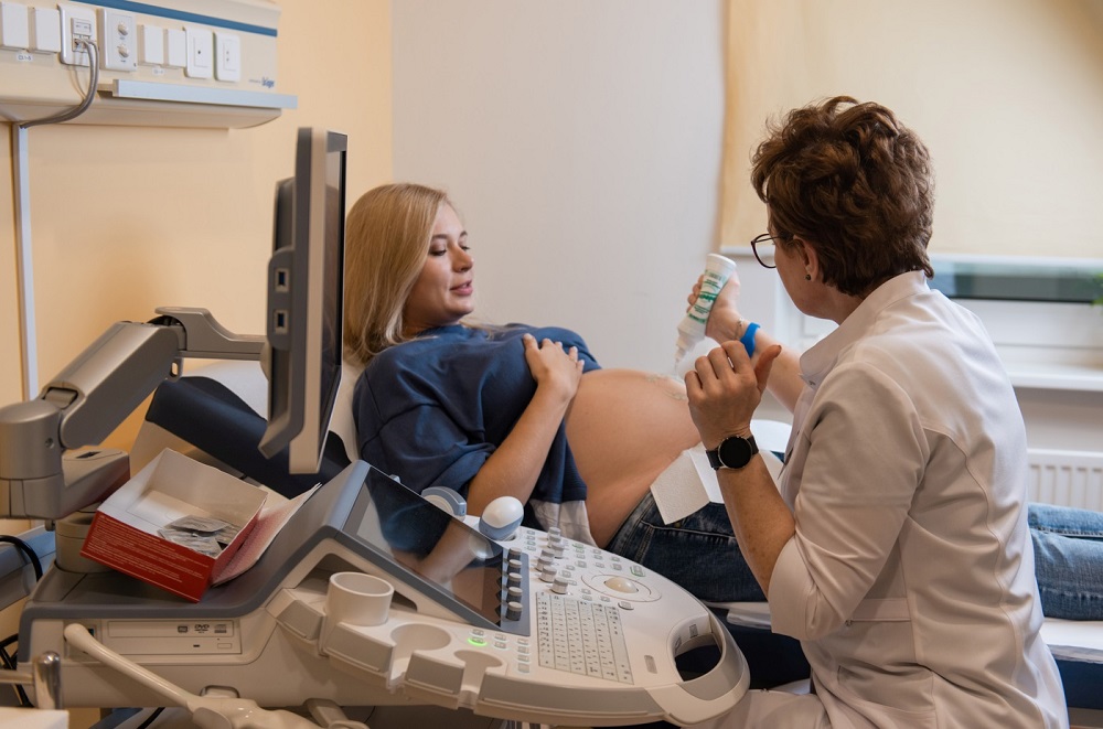
Ultrasound is a highly informative and safe diagnostic method. Today it is the most accurate diagnostic method for monitoring pregnancy. With its help, you can give an objective, competent assessment of the state of the reproductive system of a pregnant woman, monitor the correct development of the fetus, and identify pathologies (if any).

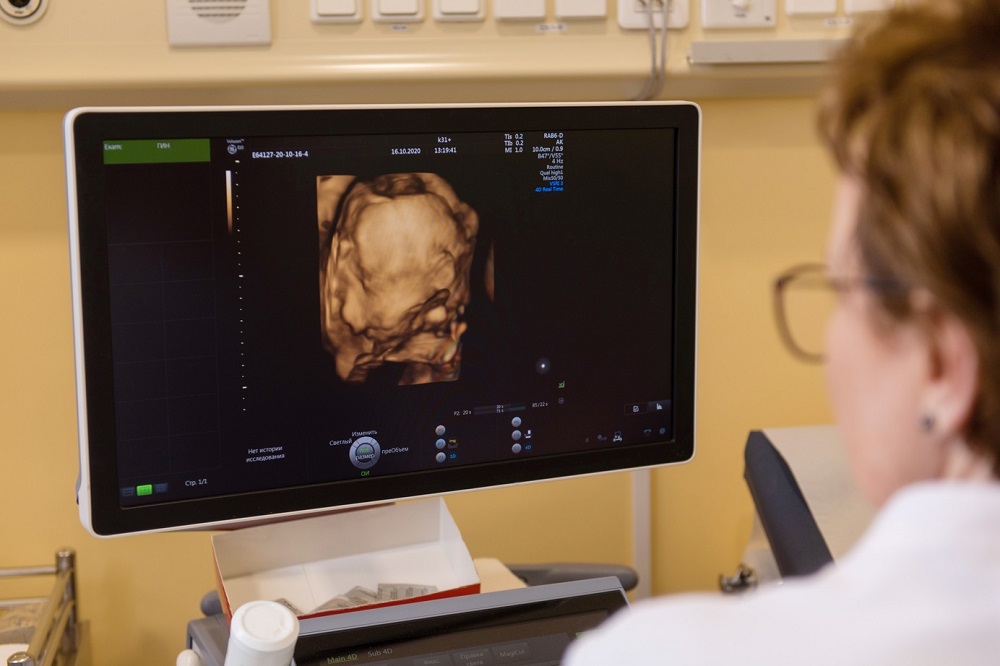
The technique allows to carry out not only two-dimensional diagnostics, but also three- and even four-dimensional research. With a two-dimensional diagnostic study, a monochrome image is obtained in only two planes. 4D ultrasound provides a color, three-dimensional image, makes it possible to see the movement of the fetus in real time.

The resulting volumetric ultrasound is not just a spectacular, detailed picture. First of all, it is a valuable tool that provides the specialist with additional information during the examination.

4D ultrasound can be performed on the recommendation of a doctor or at the request of the expectant mother. Such a study is usually prescribed for the following pathologies:

  • Suspicion of the presence of external malformations in the fetus (underdeveloped limbs, extra fingers, splitting of the upper lip, etc.).
  • Serious pathology of the placenta.
  • Incorrect development of the spine.
  • Incorrect formation of the neural tube, etc.

4D ultrasound is performed for surrogate mothers and pregnant women after IVF. It is also done at a high risk of developing a hereditary disease, the threat of miscarriage and the presence of serious pathologies in the expectant mother, requiring careful monitoring of the dynamics of fetal development.

By what indicators is 4D ultrasound superior to a standard 2D study?

  • Provides maximum information content, allows you to consider even those areas that are invisible during a two-dimensional diagnostic study.
  • Provides visualization of the smallest details, allows you to have a good look at any parts of the body - the head, face, neck, collar area, limbs, spine, anterior abdominal wall.
  • Gives minimal image error.
  • Provides timely, reliable detection of violations in the development of the fetus at any stage of pregnancy, including the earliest.
  • Provides the most accurate and informative data on the development of internal organs.
  • It makes it possible to see the appearance of the unborn child, to examine his face in detail, to take detailed photos for future parents.

When and how is the procedure carried out?

4D ultrasound is similar to standard ultrasound screening. The most suitable time for conducting a volumetric study is the period from the twentieth to the twenty-eighth week of pregnancy. At this time, you can not only determine the sex of the child, but also see the features of his face, get an idea about his appearance and the state of the internal organs.

During such an examination, the doctor gets the opportunity to check for the presence of various pathologies of pregnancy (polyhydramnios or oligohydramnios, abnormalities of the placenta, etc.). He will also be able to identify fetal pathologies (if any) at an early stage - its internal organs, joints, bones, spine, maxillofacial region. According to deviations in the appearance of the unborn child, the doctor may conclude that he has some genetic diseases, for example, Down's disease.

There are no absolute contraindications and any restrictions for 4D ultrasound, this examination is safe for pregnant women and can be performed on every pregnant patient.

During the procedure, the woman lies on her back, the study is carried out through the abdominal wall. Before the examination, the doctor applies a special medical gel to the surface of her abdomen. After that, the specialist proceeds to the procedure, the duration of which is about half an hour.

What equipment is used for diagnostics?

To conduct a volumetric study, innovative ultrasound devices VOLUSON E10 are used. This diagnostic equipment of the expert category made it possible to bring the efficiency, quality and automation of ongoing research to a fundamentally new level. A large number of software protocols, modes and additional options make it possible to use such ultrasonic devices to perform the most complex studies.

The use of premium-class expert diagnostic equipment for ultrasound screening allows for volumetric scanning, obtaining the highest quality image.

The use of this diagnostic ultrasound scanner made it possible to make communication between the doctor and the patient more effective, which led to an increase in the quality of medical services.

Service record


Specialists

All specialists
Andreytseva
Marina Igorevna

Ultrasound Diagnostic Doctor

PhD

Seleznev
Denis Evgenyevich

Head of endoscopy department, endoscopist

PhD

Sinitsyn
Valentine Evgenyevich

Chief Consultant in Radiation Diagnostics, Radiologist

Doctor of Sciences, PhD, professor

Abalmasov
Vladimir Georgievich

Leading specialist in ultrasound diagnostics

Doctor of Sciences, PhD, professor

Kondratyev
Eugene Valerevich

Leading CT Specialist, MRI

PhD, leading research fellow

Schulz
Eugene Igorevich

Leading neuroradiologist in MRI and CT diagnostics

PhD

Mebonia
Sofia Teymurazovna

Functional Diagnostic Doctor

Sokolova
Alena Victorovna

Ultrasound Diagnostic Doctor

PhD

Solovieva
Inna Borisovna

Laboratory diagnostics doctor

Malakhova
Elena Nikolaevna

Ultrasound Diagnostic Doctor

Panfilova
Ekaterina Alexandrovna

Ultrasound Diagnostic Doctor

PhD

Belevskaya
Anna Andreevna

Functional Diagnostic Doctor

Vasilieva
Yulia Nikolaevna

Radiologist, leading specialist in radiation diagnostics

PhD

Shirokih
Pavel Nikolaevich

Ultrasound and Prenatal Diagnostic Doctor

Parinova
Svetlana Vasilevna

Functional Diagnostic Doctor

Isropilova
Leila T. Alievna

Ultrasound Diagnostic Doctor

Mikula
Lada Nikolaevna

Ultrasound Diagnostic Doctor

Maso
Michael Lvovich

Mammologist, radiologist, ultrasound diagnostician

PhD

Gadzhieva
Khaybat Hadisovna

Ultrasound Diagnostic Doctor

PhD

Goryunova
Tatiana Vyacheslavovna

Cardiologist, functional diagnostics doctor

Baranova (Vorfolomeeva)
Oksana Vladimirovna

Ultrasound diagnostics doctor

First category doctor

Kozlova
Alla Valerievna

Ultrasound diagnostics doctor

Karpova
Maria Andreevna

Head of the mammological center

PhD

Samtanova
Baira Eduardovna

Doctor of clinical laboratory diagnostics

Achba
Maya Otarovna

Radiologist, ultrasound diagnostician, oncologist-mammologist

Garanina
Anna Stanislavovna

Coloproctologist, endoscopist

PhD

Puzakov
Kirill Borisovich

Leading Consultant in Radiology, Radiologist