
Gynecological ultrasound — diagnostics of pelvic organs using expert-class equipment
Gynecological ultrasound — ultrasound examination of the uterus, ovaries, fallopian tubes, and pelvic structures. This is the first line of diagnosis for most gynecological conditions: from menstrual irregularities and inflammatory processes to fibroids, cysts, and endometrial pathologies.

specialists

equipment

treatment






At K+31, gynecological ultrasound examinations are performed by specialists qualified both as obstetricians-gynecologists and ultrasound diagnostic physicians. This means that you receive not merely images with measurements, but a clinical interpretation: the doctor immediately understands what they see and is ready to answer your questions during the appointment.
Types of gynecological ultrasound examinations
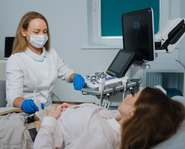
Both methods are available at K+31 — the choice depends on the clinical situation:
Transabdominal ultrasound (TA ultrasound)
The transducer is moved over the front abdominal wall. It provides an overall view of the pelvic and abdominal organs. A full bladder is required (drink 1 litre of water one hour beforehand).
Used for:
- Initial examinations of girls and virgins
- Pregnancy at any stage
- Assessment of large masses (fibroids, large cysts)
- Screening of the abdominal organs
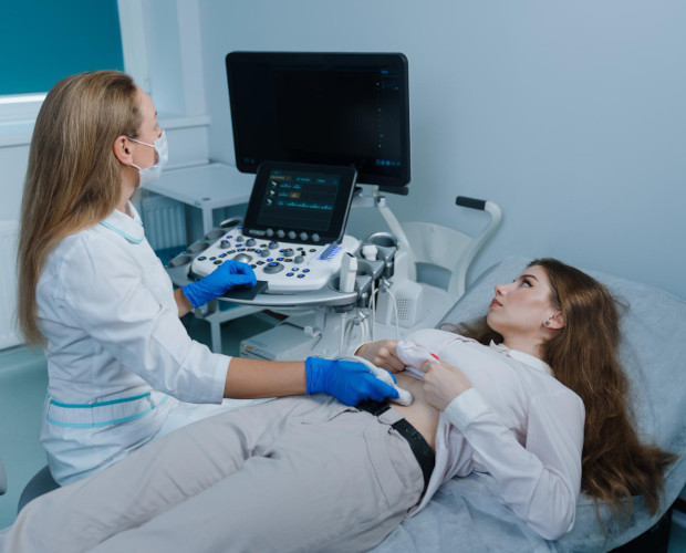
Transvaginal ultrasound (TV ultrasound)
A thin transducer is inserted into the vagina, minimizing the distance to the uterus and ovaries. Its resolution is 2–3 times higher than that of the transabdominal method. A full bladder is not required.
Used for:
- Diagnosing endometrial pathology (polyps, hyperplasia, cancer)
- Assessing the ovaries and follicular apparatus
- Early pregnancy (5–12 weeks)
- Monitoring folliculogenesis during IVF
- Identifying the source of pelvic pain
3D/4D reconstruction of the uterus
If necessary, gynecological ultrasound is supplemented with three-dimensional reconstruction. This is especially valuable for assessing the shape of the uterine cavity, diagnosing developmental anomalies (bicornuate or septate uterus), and checking IUD placement.
What does gynecological ultrasound diagnose?
Uterine pathologies:
- Uterine fibroids (subserosal, intramural, submucosal) — size, location, and effect on the cavity
- Adenomyosis — endometrial tissue growing into the muscular layer
- Endometrial polyps and hyperplasia — thickening of the lining and focal lesions
- Intrauterine adhesions (synechiae)
- Endometrial cancer — changes in the structure and thickness of the layer
- Congenital uterine anomalies
Ovarian pathologies:
- Follicular and endometriotic cysts
- Polycystic ovaries (PCOS) — a characteristic “string of pearls” appearance of small follicles
- Dermoid cysts (teratomas)
- Ovarian apoplexy and torsion — emergency conditions
- Ovarian cancer — heterogeneous structure and ascites
Inflammatory diseases:
- Endometritis — thickening and heterogeneity of the endometrium
- Salpingitis and salpingo-oophoritis — fluid in the fallopian tubes (hydrosalpinx)
- Tubo-ovarian abscess
Follow-up after interventions:
- Position of the intrauterine device (IUD)
- Condition of the scar after a caesarean section
- Retained products of conception after miscarriage or abortion
Advantages of gynecological ultrasound at K+31
- Gynecologists qualified in ultrasound diagnostics — the diagnosis is made by the specialist, not interpreted later by another doctor
- Expert-class equipment with Doppler mapping — assessment of blood flow in uterine vessels and neoplasms
- Unified medical record — ultrasound data are automatically available to the treating gynecologist at K+31
- Confidentiality — private rooms and a sensitive approach
- Appointments at convenient times — including evenings and weekends

Frequently Asked Questions (FAQ)
Which type of ultrasound is more accurate: transabdominal or transvaginal?
Transvaginal ultrasound provides considerably higher resolution for the uterus and ovaries. Transabdominal ultrasound is better suited for an overall assessment and for patients for whom transvaginal access is not possible. Most often, both methods are performed sequentially.
Is transvaginal ultrasound painful?
No. The transducer has a small diameter and the procedure is painless. If you have inflammation or experience discomfort during insertion, tell the doctor — they will adjust the pace and depth of insertion.
When is the best time to have an ultrasound if the menstrual cycle is irregular?
On days 5–7 from the start of menstruation, to assess the baseline condition of the uterus and ovaries. If your period is late, the examination can be performed on any day.
Can ultrasound determine the cause of infertility?
Ultrasound identifies anatomical causes: fibroids, adenomyosis, adhesions, polycystic ovaries, and tubal obstruction in hydrosalpinx. Functional causes require additional tests, but ultrasound is a mandatory first step.
Is ultrasound necessary if my cycle is regular and I have no symptoms?
Yes. A number of serious conditions (endometriosis, early ovarian cancer, submucosal fibroids) can remain asymptomatic for a long time. Annual preventive ultrasound is the standard of gynecological care.
Is gynecological ultrasound included in assisted reproductive technology (IVF) programmes?
Yes. During IVF preparation, gynecological ultrasound is performed repeatedly to assess the uterine cavity, monitor follicles, and provide follow-up after embryo transfer. At K+31, this programme is managed under a unified assisted reproductive technology protocol.
Is gynecological ultrasound performed for girls and adolescents?
Yes, using the transabdominal method. K+31 has a paediatric gynecologist who specializes in diagnostics for minors.
Our doctors






































This award is given to clinics with the highest ratings according to user ratings, a large number of requests from this site, and in the absence of critical violations.

This award is given to clinics with the highest ratings according to user ratings. It means that the place is known, loved, and definitely worth visiting.

The ProDoctors portal collected 500 thousand reviews, compiled a rating of doctors based on them and awarded the best. We are proud that our doctors are among those awarded.
Make an appointment at a convenient time on the nearest date
Price
Services

Appointment to the doctor




Reviews
Our clinics


Application “Personal Account K+31”











