Vascular Ultrasound
Ultrasound is a highly informative, non-invasive, portable, and painless method for visualizing human organs and tissues, widely used in all areas of modern medicine. Ultrasound waves propagate through the surrounding medium at a frequency of over 20,000 Hz. The propagation speed of the ultrasound wave is determined by the density and elasticity of the medium. The average propagation speed of ultrasound in human tissue is 1540 m/s—most ultrasound diagnostic devices are programmed for this speed.

specialists

equipment

treatment


Indications for vascular ultrasound

The examination is prescribed for the following indications:
- Headache
- Tinnitus or ringing in the ears
- Dizziness, pre-syncope
- Varicose veins
- Pain, swelling, heaviness in the legs
- Monitoring after injuries, surgeries, and procedures
Regular diagnostics of the blood vessels supplying the brain and extremities are necessary for people with hypertension, diabetes, high cholesterol, and a hereditary predisposition to cardiovascular disease. The examination not only confirms the presence of existing pathologies but also identifies diseases in the early stages, when symptoms are minimal or absent. Ultrasound is also used to monitor the effectiveness of prescribed therapy and to monitor recovery after surgery.
Contraindications
Vascular ultrasound is considered a safe diagnostic method, so there are no serious contraindications. However, there are some limitations:
- Skin damage in the area examined (infections, burns, wounds)
- Severe pain that prevents the transducer from making contact with the surface
- Acute mental disorders that prevent the patient from remaining still during the procedure
Eliminating time constraints allows the physician to perform the examination without risk to the patient, obtain accurate results, and use them for timely treatment or preventative planning.
Types of Vascular Ultrasound
The use of different types of vascular and arterial ultrasound helps detect early signs of pathologies and prevent the development of serious complications.
Vascular ultrasound is divided into three types based on the level of information content and technical capabilities:
- Doppler ultrasonography. A basic diagnostic method that evaluates the speed and direction of blood flow. It helps identify stenosis, thrombosis, and venous congestion in the extremities
- Duplex scanning. Combines standard ultrasound with Doppler ultrasonography. In addition to blood flow velocity data, the physician obtains real-time images of the vessels themselves, assessing their patency, wall thickness, and the presence of atherosclerotic plaques. It is used to evaluate the vessels of the neck and brain
- Triplex scanning. A modern and informative method that combines duplex imaging with blood flow mapping. It allows for the assessment of the vascular network, identifying thrombosis, atherosclerotic plaques, aneurysms, and stenosis
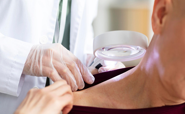
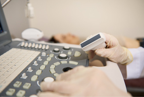
General information about the procedure
How is a vascular ultrasound performed?
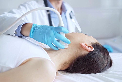
The examination is performed on an outpatient basis, in a comfortable setting. On average, the procedure takes 20-40 minutes, depending on the area being examined. Before the procedure, the patient is asked to remove jewelry and clothing in the area where the vessels will be scanned. They then lie down on a comfortable examination table for the vascular ultrasound. During the examination, the doctor may ask you to turn your head slightly or change your body position to access all the vessels. For a leg ultrasound, the examination is performed while lying on your back, or sometimes while standing, to assess venous outflow in different positions.
The doctor applies a clear gel to the skin in the area being examined, which improves the ultrasound probe's contact with the body surface and enhances image quality. The specialist then slowly moves the probe over the skin, assessing the vessels on the machine's screen. After the procedure, the gel is removed with a tissue, and the patient can immediately return to their daily activities.


Clinic Advantages
The K+31 Medical Center in Moscow employs qualified doctors with many years of experience. They use expert-grade equipment for diagnostic procedures, guaranteeing accurate examinations and accurate interpretation of results. All services are provided promptly, and interpretations are available immediately after the examination. This allows for prompt decisions regarding further treatment or preventative measures.
The clinic places special emphasis on patient comfort: modern offices, comfortable examination tables, attentive staff, and a personalized approach create the ideal conditions for examinations. The doctor explains each step of the procedure in detail, helping patients better understand the diagnostic process and reduce anxiety.
FAQ
Do I need to schedule an appointment in advance?
How long does the procedure take?
What's the difference between duplex and triplex scanning?
What's the cost?
Our doctors


This award is given to clinics with the highest ratings according to user ratings, a large number of requests from this site, and in the absence of critical violations.

This award is given to clinics with the highest ratings according to user ratings. It means that the place is known, loved, and definitely worth visiting.

The ProDoctors portal collected 500 thousand reviews, compiled a rating of doctors based on them and awarded the best. We are proud that our doctors are among those awarded.
Make an appointment at a convenient time on the nearest date
Price

Appointment to the doctor
Reviews


Our clinics

Application “Personal Account K+31”


What is vascular ultrasound?
Ultrasound diagnostics can detect narrowing or blockages of arteries and veins, circulatory disorders, blood clots, atherosclerotic plaques, and aneurysms. It is also performed before surgeries or procedures involving blood vessels.
Unlike other diagnostic methods, ultrasound does not require the administration of a contrast agent, causes no discomfort, and is suitable for patients of any age. It can be performed repeatedly without harm to health, which is important for patients with chronic diseases or after vascular interventions. The results of the examination are often used to plan further therapy and prevent stroke or thrombosis. You can have a vascular ultrasound in Moscow at the K+31 Clinic.